<acronym id="vpKZw97"></acronym>
Skip to main page content
U.S. flag

An official website of the United States government

Dot gov

The . gov means it’s official. Federal government websites often end in . gov or . mil. Before sharing sensitive information, make sure you’re on a federal government site VSports app下载. .

Https

The site is secure V体育官网. The https:// ensures that you are connecting to the official website and that any information you provide is encrypted and transmitted securely. .

. 2008 Feb 1;121(Pt 3):290-7.
doi: 10.1242/jcs.017343. Epub 2008 Jan 15.

Cytoplasmic condensation is both necessary and sufficient to induce apoptotic cell death (VSports手机版)

Affiliations

Cytoplasmic condensation is both necessary and sufficient to induce apoptotic cell death

Nola Jean Ernest et al. J Cell Sci. .

Abstract

Programmed cell death (apoptosis) is important in tissue maintenance. Hallmarks of apoptosis include caspase activation, DNA fragmentation and an overall reduction in cell volume VSports手机版. Whether this apoptotic volume decrease (AVD) is a mere response to initiators of apoptosis or whether it is functionally significant is not clear. In this study, we sought to answer this question using human malignant glioma cells as a model system. In vivo, high grade gliomas demonstrate an increased percentage of apoptotic cells as well as upregulation of death ligand receptors. By dynamically monitoring cell volume, we show that the induction of apoptosis, via activation of either the intrinsic or extrinsic pathways with staurosporine or TRAIL, respectively, resulted in a rapid AVD in D54-MG human glioma cells. This decrease in cell volume could be prevented by inhibiting the efflux of Cl(-) through channels. Such suppression of AVD also reduced the activation of caspases 3, 8 and 9 and suppressed DNA fragmentation. Importantly, experimental manipulations that reduce the cell volume to 70% of the original volume for periods of at least 3 hours were sufficient to initiate apoptosis even in the absence of death ligands. Hence, this data suggests that cell condensation is both necessary and sufficient for the induction of apoptosis. .


Figures

Fig. 1
Fig. 1
Apoptosis is initiated in human glioma cells in response to staurosporine (STS) and TRAIL treatment. (A) Caspase activity was measured in D54-MG cells treated with normal bath, 1 μM STS or 50 ng/ml TRAIL by a luminescence assay. Each time point was normalized to the luminescence of cells after 0 hours of treatment with normal bath. Data are from three experiments. Error bars represent s.e.m. and n=3 samples per experiment. (B) Western blots of cell lysates from cells treated for 1, 3 or 7 hours were probed with antibodies against the cleaved, activated forms of caspases 3, 8 and 9. (C) Representative images demonstrate DNA fragmentation (fluorescein-labeled nuclei) after treatment with normal bath, STS or TRAIL for 7 hours. Merged images show both fragmented nuclei (green) and total nuclei present by DAPI staining (blue). Bar, 100 μm.
Fig. 2
Fig. 2
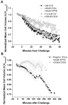
Apoptotic cell condensation is sensitive to the Cl--channel inhibitor DIDS. (A,B) Normalized mean cell volumes (MCVs) of D54-MG were recorded following addition of STS (A) or TRAIL (B) in the presence of the Cl--channel inhibitors NPPB, DIDS or Cd2+ or the KCC inhibitor DIOA. Data are the mean results from three independent experiments. Error bars represent s.e.m. with n=10,000-20,000 cells.
Fig. 3
Fig. 3
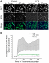
Apoptosis initiation results in DIDS-sensitive Cl- movement across the plasma membrane. (A) Representative images depict cell size as well as SPQ and GFP intensity just prior to (t=0) and 27 minutes after the addition of control or STS media. (B) Mean time normalized intensity curves quantify the changes depicted in A. Data are the mean results from three independent experiments. Error bars represent s.e.m. with n=10 samples in each experiment.
Fig. 4
Fig. 4
DNA fragmentation and caspase activation are also sensitive to DIDS inhibition of AVD. (A) Representative images demonstrate the presence or absence of DNA fragmentation (FITC-labeled nuclei) in cells treated for 7 hours with STS or TRAIL in the presence or absence of DIDS. Merged images show fragmented nuclei (green) as well as DAPI staining of all nuclei present (blue). (B) Western blots of lysates from similarly treated cells were probed with antibodies to activated, cleaved caspases 3, 8 and 9. Actin was used as a loading control.
Fig. 5
Fig. 5
DIDS inhibition of AVD has variable effects on cell viability. Percentage cell viability as measured by Trypan Blue exclusion after application of STS or TRAIL for 7 hours. *P<0.05 compared with untreated control cells.
Fig. 6
Fig. 6
Hyperosmotic cell shrinkage results in activation of caspase 3. (A) Normalized MCVs of D54-MG cells were recorded following the addition of 150 or 300 mOsm mannitol in the presence or absence of 2 mM glutamine. Data are the mean results from three independent experiments. Error bars represent s.e.m. with n=10,000-20,000 cells. (B) Caspase 3 activity, as measured by luminescence, in cells exposed to isosmotic (300 mOsm) or hyperosmotic (337.5-600 mOsm) bath solutions for 24 hours. Activity normalized to 300 mOsm exposure. Data are from three experiments. Error bars represent s.e.m. and n=3 samples per experiment. *P<0.05.
Fig. 7
Fig. 7
The ‘time threshold’ to activate is reduced with a greater cell shrinkage. Cells were exposed to bath solution made hyperosmotic by the addition of NaCl (final osmolarity 450 or 600) and were then ‘rescued’ from shrinkage by the addition of ddH2O after 30 minutes, 1, 4 or 8 hours. (A) Normalized MCVs of D54-MG cells during exposure to and rescue from hypertonic challenge in a representative experiment. (B) Caspase 3 activity as measured by luminescence. Activity is normalized to 300 mOsm (no challenge) exposure. Data are from three independent experiments. Error bars represent s.e.m. and n=3 samples per experiment. *P<0.05 compared to unchallenged control cells (not shown).
Fig. 8
Fig. 8
Proposed model for the initiation of apoptosis by cell shrinkage. Cell shrinkage may result in the increased concentration of auto-activating enzymes (top panel) or the increase in proximity of cell surface receptors (bottom panel). The activation of such enzymes/receptors may result in the initiation of apoptosis in the absence of other drugs or the acceleration of the pathway in the presence of other apoptosis initiators.

"V体育平台登录" References

    1. Bortner CD, Cidlowski JA. Absence of volume regulatory mechanisms contributes to the rapid activation of apoptosis in thymocytes. Am. J. Physiol. 1996;271:C950–C961. - PubMed
    1. d’Anglemont de Tassigny A, Ghaleh B, Souktani R, Henry P, Berdeaux A. Hypo-osmotic stress inhibits doxorubicin-induced apoptosis via a protein kinase A-dependent mechanism in cardiomyocytes. Clin. Exp. Pharmacol. Physiol. 2004;31:438–443. - PubMed
    1. Ernest NJ, Sontheimer H. Extracellular glutamine is a critical modulator for regulatory volume increase in human glioma cells. Brain Res. 2007;1144:231–238. - PMC - PubMed
    1. Ernest NJ, Weaver AK, Van Duyn LB, Sontheimer HW. Relative contribution of chloride channels and transporters to regulatory volume decrease in human glioma cells. Am. J. Physiol. Cell Physiol. 2005;288:1451–1460. - PMC - PubMed
    1. Fumarola C, Zerbini A, Guidotti GG. Glutamine deprivation-mediated cell shrinkage induces ligand-independent CD95 receptor signaling and apoptosis. Cell Death Differ. 2001;8:1004–1013. - PubMed

Publication types

MeSH terms

VSports在线直播 - LinkOut - more resources