Skip to main page content
U.S. flag

An official website of the United States government

Dot gov

The . gov means it’s official. Federal government websites often end in . gov or . mil. Before sharing sensitive information, make sure you’re on a federal government site. VSports app下载.

Https

The site is secure. The https:// ensures that you are connecting to the official website and that any information you provide is encrypted and transmitted securely. V体育官网.

. 2007 Nov 19;97(10):1381-7.
doi: 10.1038/sj.bjc.6604059. Epub 2007 Oct 30.

Photodynamic therapy-generated vaccines: relevance of tumour cell death expression (V体育安卓版)

Affiliations

Photodynamic therapy-generated vaccines: relevance of tumour cell death expression

M Korbelik et al. Br J Cancer. .

Abstract

Recent investigations have established that tumour cells treated in vitro by photodynamic therapy (PDT) can be used for generating potent vaccines against cancers of the same origin. In the present study, cancer vaccines were prepared by treating mouse SCCVII squamous cell carcinoma cells with photosensitiser chlorin e6-based PDT and used against poorly immunogenic SCCVII tumours growing in syngeneic immunocompetent mice. The vaccine potency increased when cells were post-incubated in culture after PDT treatment for 16 h before they were injected into tumour-bearing mice. Interfering with surface expression of phosphatidylserine (annexin V treatment) and apoptosis (caspase inhibitor treatment) demonstrated that this post-incubation effect is affiliated with the expression of changes associated with vaccine cell death. The cured mice acquired resistance to re-challenge with the same tumour, while the engagement of cytotoxic T lymphocytes was demonstrated by detection of high numbers of degranulating CD8+ cells in vaccinated tumours. The vaccines prepared from ex vivo PDT-treated SCCVII tumour tissue were also highly effective, implying that surgically removed tumour tissue can be directly used for PDT vaccines. This opens attractive prospects for employing PDT vaccines tailored for individual patients targeting specific antigens of the patient's tumour VSports手机版. .


Figures
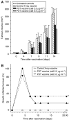
Figure 1
Figure 1
The effect of photodynamic therapy (PDT)-generated vaccine prepared using photosensitiser ce6 on growth of established SCCVII tumours. The vaccine was prepared by standard vaccine protocol (see Materials and Methods section) involving the incubation of SCCVII cells with ce6 (0.5 or 2.5 μg ml−1) for 30 min, then illumination (1 J cm−2), and exposure to X-rays (60 Gy) followed immediately by their injection in SCCVII tumour-bearing mice (2 × 10⁷ cells per mouse, peritumorally). The therapy response was monitored by tumour size measurement and is presented as (A) means for tumour volume values plus s.d., and (B) percentage of growth-inhibited tumours (smaller than the means minus two-fold s.d. of unvaccinated control group). The controls are showing growth of vaccine untreated and X-ray only vaccine treated tumours. Each treatment group consisted of six mice. *Indicates statistical significance (P<0.05) for the difference in tumour growth compared to untreated controls.
Figure 2
Figure 2
Comparison of the effects of photodynamic therapy (PDT) vaccines prepared by standard and post-incubation protocols. The preparation of PDT vaccine was based on the treatment of SCCVII cells with ce6 (0.5 μg ml−1) as described for Figure 1, except that 16-h post PDT incubation was or was not also included (see Materials and Methods section). The response of SCCVII tumours after peritumoral injection of 2 × 10⁷ cells per mouse is presented as survival of mice (terminating when tumours reached 100 mm3). Each treatment group consisted of six mice. *Indicates statistical significance (P<0.05) for the difference in survival compared to untreated controls; **Indicates statistical significance (P<0.05) for the difference in survival compared to the vaccine with no post-incubation treatment group.
Figure 3
Figure 3
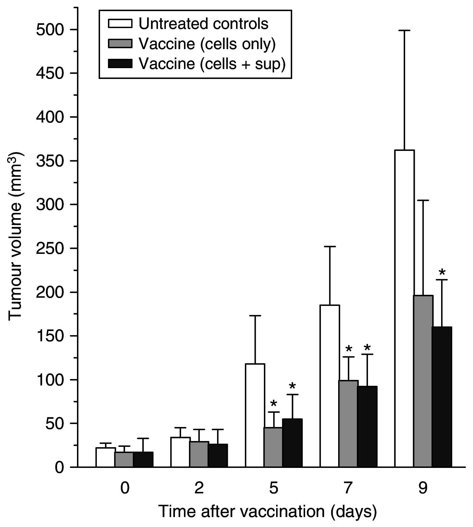
Testing of photodynamic therapy (PDT) vaccine cell supernatant. The PDT vaccine was prepared using the post-incubation protocol described for Figure 2. In addition to vaccine cells, the vaccine injection in one group of mice included concentrated post-incubation medium supernatants. The results are presented as histograms depicting means for tumour volume plus s.d. Each treatment group consisted of six mice. *Indicates statistical significance (P<0.05) for the difference in tumour size compared to the untreated controls at the same time interval.
Figure 4
Figure 4
The effect of exposing photodynamic therapy (PDT) vaccine cells to annexin V. Mice-bearing SCCVII tumours received peritumoral injection of PDT vaccine consisting of SCCVII cells treated by the post-incubation protocol described for Figure 2, but including in one group also a 30-min exposure of cells to annexin V (0.2 mg ml−1, see Materials and Methods section). The therapy response was monitored by tumour size measurement and is presented as percentage of growth-inhibited tumours (same as Figure 1B). Each treatment group consisted of six mice. *Indicates statistical significance (P<0.05) for the difference in tumour growth compared to untreated controls; **Indicates statistical significance (P<0.05) for the difference in tumour growth compared to the annexin V-modified vaccine treatment group.
Figure 5
Figure 5
The effect of apoptosis inhibition in photodynamic therapy (PDT) vaccine cells. Mice-bearing SCCVII tumours received peritumoral injection of PDT vaccine consisting of SCCVII cells treated by the post-incubation protocol described for Figure 2, except that Z-DEVD (33.4 μg ml−1) was present in the post-incubation medium for one group. The therapy response was monitored by tumour size measurement and is presented as the means for tumour volume values plus s.d. Each treatment group consisted of six mice. *Indicates statistical significance (P<0.05) for the difference in tumour size compared to the untreated controls at the same time interval; #Indicates statistical significance (P<0.05) for the difference in tumour size compared to the vaccine without Z-DEVD treatment group at the same time interval. Φ=66.7% were tumour free at 90 days post-vaccination.
Figure 6
Figure 6
Detection of degranulating cytotoxic T lymphocytes in tumours treated by photodynamic therapy (PDT) vaccine. Mice-bearing SCCVII tumours received peritumoral injection of PDT vaccine (SCCVII cells treated by the post-incubation protocol described for Figure 2). The tumours were excised 3 days later and disaggregated into single cell suspensions that were stained with antibodies against mouse CD8, CD11b and CD107a antigens, and analysed by flow cytometry. Degranulating cytotoxic T lymphocytes were identified as CD8+CD107a+ cells in populations gated negative for staining with myeloid marker CD11b. Samples from tumours that regressed by the time of excision (good early responders) and those from tumours that continued growing (poor early responders) were analysed separately. Also included were samples from control sham-vaccinated tumours (saline injected) and from tumours injected with SCCVII cells treated only with X-rays. The experimental groups consisted of four or five mice. Bars are s.d. *Indicates statistical significance (P<0.05) for the difference in the number of CD8+CD107a+ cells compared to the untreated control group; **Indicates statistical significance (P<0.05) for the difference in the number of CD8+CD107a+ cells compared to the poor early responders group; #Indicates statistical significance (P<0.05) for the difference in the number of CD8+CD107a+ cells compared to the control X-ray vaccine group.
Figure 7
Figure 7
Comparison of the effects of locally (peritumorally) or distally administered photodynamic therapy (PDT) vaccines. Mice-bearing SCCVII tumours received PDT vaccine (prepared by the post-incubation protocol described for Figure 2) injected either peritumorally or at a distal location (subcutaneously at dorsal neck site). Tumour responses are presented as percentage of growth-inhibited tumours (same as in Figure 1B). Each treatment group consisted of six mice. *Indicates statistical significance (P<0.05) for the difference in tumour growth compared to the untreated controls group; **Indicates statistical significance for the difference in tumour growth compared to the distal vaccine treatment group.
Figure 8
Figure 8
The effect of photodynamic therapy (PDT) vaccine prepared directly from tumour tissue. Mice-bearing SCCVII tumours received a peritumoral injection of PDT vaccine prepared either from SCCVII cells or from SCCVII tumour tissue brei using in both cases the post-incubation protocol described for Figure 2). Tumour responses are presented as percentage of growth-inhibited tumours (same as in Figure 1B). Each treatment group consisted of six mice. *Indicates statistical significance (P<0.05) for the difference in tumour growth compared to the untreated controls group.

References

    1. Betts MR, Brenchley JM, Price DA, De Rosa SC, Douek DC, Roederer M, Koup RA (2003) Sensitive and viable identification of antigen-specific CD8+ T cells by flow cytometric assay for degranulation. J Immunol Methods 281: 65–78 - PubMed
    1. Brown SB, Brown EA, Walker I (2004) The present and future role of photodynamic therapy in cancer treatment. Lancet Oncol 5: 497–508 - PubMed
    1. Burkett MW, Shafer-Weaver KA, Strobl S, Baseler M, Malyguine A (2005) A novel flow cytometric assay for evaluating cell-mediated cytotoxicity. J Immunother 28: 396–402 - PubMed
    1. Castano AP, Mroz P, Hamblin MR (2006) Photodynamic therapy and anti-tumour immunity. Nat Rev Cancer 6: 535–545 - PMC - PubMed
    1. Copier J, Dalgleish A (2006) Overview of tumour cell-based vaccines. Int Rev Immunol 25: 297–319 - PubMed

Publication types

MeSH terms

LinkOut - more resources