Skip to main page content
U.S. flag

An official website of the United States government

Dot gov

The VSports app下载. gov means it’s official. Federal government websites often end in . gov or . mil. Before sharing sensitive information, make sure you’re on a federal government site. .

Https

The site is secure. The https:// ensures that you are connecting to the official website and that any information you provide is encrypted and transmitted securely. V体育官网.

. 2008 Jan;76(1):403-16.
doi: 10.1128/IAI.01189-07. Epub 2007 Oct 29.

VSports最新版本 - Host transmission of Salmonella enterica serovar Typhimurium is controlled by virulence factors and indigenous intestinal microbiota

Affiliations

"VSports最新版本" Host transmission of Salmonella enterica serovar Typhimurium is controlled by virulence factors and indigenous intestinal microbiota

V体育2025版 - Trevor D Lawley et al. Infect Immun. 2008 Jan.

"V体育官网" Abstract

Transmission is an essential stage of a pathogen's life cycle and remains poorly understood. We describe here a model in which persistently infected 129X1/SvJ mice provide a natural model of Salmonella enterica serovar Typhimurium transmission. In this model only a subset of the infected mice, termed supershedders, shed high levels (>10(8) CFU/g) of Salmonella serovar Typhimurium in their feces and, as a result, rapidly transmit infection. While most Salmonella serovar Typhimurium-infected mice show signs of intestinal inflammation, only supershedder mice develop colitis. Development of the supershedder phenotype depends on the virulence determinants Salmonella pathogenicity islands 1 and 2, and it is characterized by mucosal invasion and, importantly, high luminal abundance of Salmonella serovar Typhimurium within the colon VSports手机版. Immunosuppression of infected mice does not induce the supershedder phenotype, demonstrating that the immune response is not the main determinant of Salmonella serovar Typhimurium levels within the colon. In contrast, treatment of mice with antibiotics that alter the health-associated indigenous intestinal microbiota rapidly induces the supershedder phenotype in infected mice and predisposes uninfected mice to the supershedder phenotype for several days. These results demonstrate that the intestinal microbiota plays a critical role in controlling Salmonella serovar Typhimurium infection, disease, and transmissibility. This novel model should facilitate the study of host, pathogen, and intestinal microbiota factors that contribute to infectious disease transmission. .


Figures

FIG. 1.
FIG. 1.
Transmission of Salmonella serovar Typhimurium between hosts results in a systemic infection. Four orogastrically infected donor mice shedding Salmonella serovar Typhimurium were housed with one uninfected mouse. (a) Fecal shedding was monitored every 1 to 3 days by culturing fresh fecal pellets from the previously uninfected mouse (asterisks) and donor mice (gray circles) for Salmonella serovar Typhimurium. (b) Feces from the newly infected mouse (asterisks) and orogastrically infected mice (gray circles) were monitored for the induction of Salmonella serovar Typhimurium-specific IgA during infection. (c) Levels of Salmonella serovar Typhimurium within the cecum, Peyer's patches (PP), mLN, spleen, and liver were determined for the originally uninfected mouse (asterisks) and the orogastrically infected mice (gray circles). The detection limit for each tissue is indicated by the dashed line. (d) Titers of Salmonella serovar Typhimurium-specific IgG in the serum of mice, showing comparable levels in orogastrically (gray bars) and transmission-infected (black bar) mice.
FIG. 2.
FIG. 2.
Transmission of Salmonella serovar Typhimurium between hosts requires high levels of shedding which is dependent on SPI1 and SPI2. (a and b) Mice were infected orogastrically with 108 CFU of either wild-type (wt) (n = 45), sipB::Cm (n = 30), or ssaV::Kan (n = 30) Salmonella serovar Typhimurium (Stm), and the peak fecal shedding levels (a) and duration of fecal shedding (b) were monitored. (c) Summary of single-donor transmission experiments in which one infected mouse was housed with four uninfected mice for 1 to 58 days and shedding was monitored. The peak level of shedding from donor mice was plotted against the percentage of uninfected mice that became infected. The shedding ranges and numbers of donor mice are indicated below the x axis. (d) A supershedder (donor) (black line) was added to a cage with four uninfected mice (gray lines) for 16 h and then removed. Shedding from the uninfected mice was monitored for 19 days. Previously uninfected mice were colonized in the liver, spleen, and mLN. The dashed line indicates the supershedding level.
FIG. 3.
FIG. 3.
SPI1 and SPI2 contribute to high levels of shedding and systemic colonization. (a to c) Representative shedding patterns for mice infected orogastrically with 108 CFU of wild-type (n = 5) (a), sipB::Cm (n = 5) (b), or ssaV::Kan (n = 5) (c) Salmonella serovar Typhimurium. The level of detection was ∼10 CFU of Salmonella/g of feces. The dashed lines at 104 and 108 CFU/g indicate the shedding categories. (d) Systemic colonization levels for the mice infected for 30 days with wild-type (wt), sipB::Cm, and ssaV::Kan Salmonella serovar Typhimurium. The colonization levels for individual mice are indicated by circles, and the geometric mean for each group is indicated by a solid line. The detection limit for each tissue is indicated by a dashed line. The P values obtained with a Mann-Whitney test comparing the geometric means for the wild-type and sipB::Cm Salmonella serovar Typhimurium strains are as follows: 0.3095 for the liver; 0.0952 for the spleen; and 0.0556 for the mLN. The P values obtained with a Mann-Whitney test comparing the geometric means for the wild-type and ssaV::Kan Salmonella serovar Typhimurium strains are as follows: 0.0079 for the liver; 0.0556 for the spleen; and 0.0177 for the mLN. The data are representative of three experiments.
FIG. 4.
FIG. 4.
Colonization by Salmonella serovar Typhimurium leads to moderate to severe colitis in supershedder mice. (a and b) Ceca of moderate-shedder mice (a) and supershedder mice (b) infected with Salmonella serovar Typhimurium for 20 days. (c) Cecum of an uninfected mouse shown for comparison. (d and e) Colons of moderate-shedder mice (d) and supershedder mice (e) infected with Salmonella serovar Typhimurium for 20 days. (f) Colon of an uninfected mouse shown for comparison. All sections were stained with hematoxylin and eosin. Scale bars = 100 μm.
FIG. 5.
FIG. 5.
Distribution of Salmonella serovar Typhimurium within colons of infected mice: levels of Salmonella serovar Typhimurium in the luminal contents and colons of supershedder (n = 4) and moderate-shedder (n = 4) mice after incubation in the absence (gray bars) and presence (open bar) of gentamicin. The detection limit was 102 CFU/g. The data are representative of three experiments.
FIG. 6.
FIG. 6.
Colons of supershedder mice contain intracellular (CD11c+ dendritic cells) and extracellular luminal Salmonella serovar Typhimurium. (a to f) Immunofluorescent photomicrographs of serial sections of colons from uninfected (a and d), moderate-shedder (b and e), and supershedder (c and f) mice. Host actin (blue) was stained with Phalloidin, and host leukocytes (red) were stained with CD11c (a, b, and c) and Gr-1 (d, e, and f) antibodies. Scale bars = 100 μm. (g and h) Immunofluorescent photomicrographs of supershedder colon stained for CD11c (red), Salmonella (green), and host nuclear DNA (blue). Scale bars = 100 μm (g) and 10 μm (h). (h) Magnified section of panel g, showing Salmonella serovar Typhimurium within a CD11c+ host cell. The XZ (above panel h) and YZ (to the right of panel h) images are sections from a three-dimensional reconstruction of the image in panel g. (i to k) Immunofluorescent photomicrographs of colonic contents from uninfected (i), moderate-shedder (j), and supershedder (k) mice stained for Gr-1 (red), Salmonella (green), and DNA (host cell or bacterial) (blue). Scale bars = 20 μm. The images are representative of the results of a microscopy analysis of tissues from five different mice.
FIG. 7.
FIG. 7.
Immunosuppression does not induce the supershedder phenotype in mice. (a) Mice were treated with dexamethasone (black bars) (n = 9) or were not treated (gray bars) (n = 9) and then were infected with Salmonella serovar Typhimurium, and shedding was monitored for 10 to 14 days to identify the peak shedding range. (b) Systemic colonization for infected mice that were treated with dexamethasone (DXM) (from the experiment whose results are shown in panel a) or were not treated (from the experiment whose results are shown in panel a). The colonization levels of individual mice are indicated by black (dexamethasone treated) or gray (untreated) circles, and the geometric means are indicated by solid lines. The detection limit for Salmonella serovar Typhimurium for each tissue is indicated by a dashed line. The levels of Salmonella serovar Typhimurium for mice treated with dexamethasone are significantly higher that the levels of Salmonella serovar Typhimurium for untreated mice as determined by the Mann-Whitney test (for the liver, P = 0.0286; for the spleen, P = 0.0286; for the mLN, P = 0.0286). (c) Mice persistently infected with Salmonella serovar Typhimurium for 110 days were treated with dexamethasone (arrow), and shedding was monitored for 21 days. (d) Systemic colonization in persistently infected mice that were treated with dexamethasone (from the experiment whose results are shown in panel c) or were not treated (data not shown). The levels of Salmonella serovar Typhimurium for mice treated with dexamethasone are significantly higher than the levels of Salmonella serovar Typhimurium for untreated mice as determined by the Mann-Whitney test (for the liver, P = 0.0079; for the spleen, P = 0.0079; for the mLN, P = 0.0079). The data are representative of two experiments.
FIG. 8.
FIG. 8.
Induction of the supershedder phenotype following a reduction in the size of the intestinal bacterial community due to streptomycin treatment. (a) Shedding patterns of mice (n = 5) that were infected for 23 days and then treated orally with 5 mg of streptomycin (arrow). (b) Q-PCR of total bacterial rRNA gene (open bars) and Salmonella serovar Typhimurium DNA (gray bars) from feces of mice prior to and after streptomycin treatment of mice that were shedding low levels of Salmonella serovar Typhimurium. The number of CFU of Salmonella serovar Typhimurium (black bars) was determined by culturing organisms from feces to compare the quantification methods. The number of CFU was not determined for the 6-h time point. The dashed line at 108 CFU/g indicates the supershedder level. The data are representative of three experiments.
FIG. 9.
FIG. 9.
Reactivation of the supershedder phenotype in chronically infected mice after the size of the indigenous microbiota was reduced by streptomycin treatment. (a) Mice chronically infected for 125 days and treated with streptomycin (arrow) (n = 10). (b) Mice chronically infected for 125 days and mock treated with PBS (arrow) (n = 10). (c) Colonization levels for various organs from mice that were treated with streptomycin or PBS. Black (streptomycin) and gray (PBS) circles indicate the levels for individual mice, and the geometric means are indicated by solid lines. The detection limit for Salmonella serovar Typhimurium for each tissue is indicated by a dashed line. The levels of Salmonella serovar Typhimurium for mice treated with streptomycin are significantly higher than the levels of Salmonella serovar Typhimurium for mock-treated mice as determined by the Mann-Whitney test (for the cecum, P = 0.029; for the colon, P = 0.029; for the feces; P = 0.029; for the liver, P = 0.029; for the spleen, P = 0.029; for the mLN, P = 0.029). (d) Levels of serum anti-Salmonella serovar Typhimurium IgG for mice treated with streptomycin or PBS. The positive control (pos.) was a mouse orally infected with 108 CFU Salmonella serovar Typhimurium for 30 days, and the negative control (neg.) was an uninfected mouse.
FIG. 10.
FIG. 10.
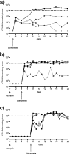
Antibiotic treatment induces a lasting predisposition to the supershedder phenotype. (a) Shedding from mice infected with 108 CFU of Salmonella serovar Typhimurium by oral gavage (n = 4). (b) Shedding from mice infected with 108 CFU of Salmonella serovar Typhimurium by oral gavage 4 days after neomycin treatment (n = 4). (c) Shedding from mice infected with 108 CFU of Salmonella serovar Typhimurium by oral gavage 7 days after neomycin treatment (n = 4). The dashed lines at 108 CFU/g indicate the supershedder level. The time of neomycin treatment on day 0 and the time of Salmonella serovar Typhimurium infection are indicated by arrows. The black lines indicate data for mice that are supershedders, whereas the data for moderate shedders are indicated by gray lines.

References

    1. Amann, R. I., B. J. Binder, R. J. Olson, S. W. Chisholm, R. Devereux, and D. A. Stahl. 1990. Combination of 16S rRNA-targeted oligonucleotide probes with flow cytometry for analyzing mixed microbial populations. Appl. Environ. Microbiol. 561919-1925. - PMC - PubMed
    1. Barthel, M., S. Hapfelmeier, L. Quintanilla-Martinez, M. Kremer, M. Rohde, M. Hogardt, K. Pfeffer, H. Russmann, and W. D. Hardt. 2003. Pretreatment of mice with streptomycin provides a Salmonella enterica serovar Typhimurium colitis model that allows analysis of both pathogen and host. Infect. Immun. 712839-2858. - PMC - PubMed
    1. Bauer-Garland, J., J. G. Frye, J. T. Gray, M. E. Berrang, M. A. Harrison, and P. J. Fedorka-Cray. 2006. Transmission of Salmonella enterica serotype Typhimurium in poultry with and without antimicrobial selective pressure. J. Appl. Microbiol. 1011301-1308. - PubMed
    1. Bing, F. C., and L. B. Mendel. 1931. The relationship between food and water intakes in mice. Am. J. Physiol. 98169-179.
    1. Brinig, M. M., P. W. Lepp, C. C. Ouverney, G. C. Armitage, and D. A. Relman. 2003. Prevalence of bacteria of division TM7 in human subgingival plaque and their association with disease. Appl. Environ. Microbiol. 691687-1694. - PMC - PubMed

Publication types

"VSports注册入口" Substances