Skip to main page content
U.S. flag

An official website of the United States government

Dot gov

The . gov means it’s official VSports app下载. Federal government websites often end in . gov or . mil. Before sharing sensitive information, make sure you’re on a federal government site. .

Https

The site is secure V体育官网. The https:// ensures that you are connecting to the official website and that any information you provide is encrypted and transmitted securely. .

. 2007 Oct 29;204(11):2641-53.
doi: 10.1084/jem.20070458. Epub 2007 Oct 8.

V体育ios版 - Cross-presentation of glycolipid from tumor cells loaded with alpha-galactosylceramide leads to potent and long-lived T cell mediated immunity via dendritic cells

Affiliations

Cross-presentation of glycolipid from tumor cells loaded with alpha-galactosylceramide leads to potent and long-lived T cell mediated immunity via dendritic cells

Kanako Shimizu et al. J Exp Med. .

Abstract

We report a mechanism to induce combined and long-lived CD4(+) and CD8(+) T cell immunity to several mouse tumors. Surprisingly, the initial source of antigen is a single low dose of tumor cells loaded with alpha-galactosylceramide (alpha-GalCer) glycolipid (tumor/Gal) but lacking co-stimulatory molecules. After tumor/Gal injection intravenously (i. v. ), innate NKT and NK cells reject the tumor cells, some of which are taken up by dendritic cells (DCs). The DCs in turn cross-present glycolipid on CD1d molecules to NKT cells and undergo maturation. For B16 melanoma cells loaded with alpha-GalCer (B16/Gal), interferon gamma-producing CD8(+) T cells develop toward several melanoma peptides, again after a single low i. v. dose of B16/Gal. In all four poorly immunogenic tumors tested, a single dose of tumor/Gal i. v VSports手机版. allows mice to become resistant to tumors given subcutaneously. Resistance requires CD4(+) and CD8(+) cells, as well as DCs, and persists for 6-12 mo. Therefore, several immunogenic features of DCs are engaged by the CD1d-mediated cross-presentation of glycolipid-loaded tumor cells, leading to particularly strong and long-lived adaptive immunity. .


Figures

Figure 1.
Figure 1.
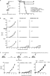
Vaccination with B16/Gal induces T cell–dependent antitumor resistance. (A) 106 α-GalCer–loaded DCs per mouse were administered to mice. 105 B16 melanoma tumor cells were injected s.c. 2 wk later. Measurement of tumor size was done at the indicated time points (n = 5 per group). Similar results were obtained in two independent experiments. (B) 105 B16/Gal or nonloaded B16 melanoma cells per mouse were administered s.c., and tumor size was followed (n = 5 per group). Similar results were obtained in two independent experiments. (C and D, left) 5 × 10⁵ α-GalCer–loaded B16 or CD1dhi-B16 tumor cells per mouse were given i.v., and 2 wk later mice were challenged with 105 parental B16 tumors s.c. (n = 10 per group). Similar results were obtained in two independent experiments. (D, right) At 2 wk after vaccination of CD4−/−, CD8−/−, and MHC II−/− mice with 5 × 10⁵ CD1dhi-B16/Gal i.v., the mice were challenged s.c. with B16 tumor cells (n = 5 per group). Similar results were obtained in two independent experiments. (E) CD19+ B cells were isolated with magnetic beads from the spleen, coated with α-GalCer, and injected together with irradiated CD1dhi-B16 cells (n = 5 per group). Data are means ± SEM. Similar results were obtained in two independent experiments.
Figure 2.
Figure 2.
Vaccination with B16-GalCer tumor cells i.v. leads to T cell immunity to several defined melanoma differentiation antigens. (A) Mice were injected i.v. with 5 × 10⁵ B16, B16/Gal, CD1dhi-B16, or CD1dhi-B16/Gal. To monitor antigen-specific T cell responses 1 and 4 wk later, CD8+ T cells were positively selected from the spleen and were cultured with splenic CD11c+ DCs from naive mice for 36 h. The DCs had been cultured for 2 h in the presence or absence of 10 μM gp10025–33, Trp2180–188, Tyrp455–463, Tyrp522–529, or Dct363–371 peptides. T cells responding to the DCs were detected by ELISPOT assay for IFN-γ production. All data are means ± SEM obtained from three independent experiments with two mice per group. *, P < 0.05 for B16/G versus CD1dhi-B16/G. (B) As in A, but the immune responses were monitored with intracellular cytokine staining. CD8+ T cells were magnetically isolated from the spleen 7 d after i.v. CD1dhi-B16/Gal. The CD8+ T cells were co-cultured for 16 h with splenic CD11c+ DCs from naive mice in the presence of brefeldin A, which had been pulsed with 10 μM Trp2180–188 peptide for 2 h, for the last 10 h. Numbers indicate the percentage of total CD8+ cells. The data are representative of three independent experiments (n = 4 per group). (C) Mice were immunized with 5 × 10⁵ CD1dhi-B16/Gal and depleted NK1.1+ cells by injections of 300 μg of anti-NK1.1(PK136) antibody per mouse at days −1, 3, and 5 (before vaccination), or days 3 and 5 (after vaccination). As in A, the TRP2-specific CD8+ T cell response was detected by ELISPOT assay for IFN-γ at day 7. Data are means ± SEM obtained from three independent experiments (n = 4 per group). **, P < 0.01.
Figure 3.
Figure 3.
Vaccination with live and irradiated tumor/Gal induces antitumor protection against several mouse tumors. (A, left) Immunization experiments were set up as shown; i.e., mice were vaccinated with 5 × 10⁶ of different forms of J558 myeloma cells, challenged at 4 wk with 106 live J558 s.c., and evaluated for their survival (n = 10 per group). Similar results were obtained in two independent experiments. (B) Mice were injected i.v. with 5 × 10⁵ live and irradiated CD1dhi-B16/Gal (n = 9 and 6 per group, respectively), 2 × 10⁶ CD1dhi-WEHI-3B/Gal (n = 5 per group), or 5 × 10⁵ CD1dhi-EL4/Gal (n = 8 and 5 per group, respectively). The mice were challenged s.c. with parental tumor cells (5 × 10⁴ B16, 2 × 10⁵ WEHI-3B, or 2 × 10⁵ EL4) 4 wk later. Tumor sizes were measured at the indicated time points. Similar results were obtained in two independent experiments. (C) Mice were immunized and challenged as indicated in the diagram (n = 5 B16 or EL4; n = 9 tumor/Gal-B16 −EL4). Data are means ± SEM. Similar results were obtained in two independent experiments.
Figure 4.
Figure 4.
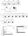
DCs are less effective than tumor/Gal in presenting OVA antigen when injected via the i.v. route. (A) Mice were adoptively transferred with 2 × 10⁶ CFSE-labeled OT-I cells and were immunized the next day with EL4(OVA) or CD1dhi-EL4(OVA) ± Gal. 3 d later, the animals were tested for proliferation of transferred OT-I cells as well as CD25 (top). Data are representative of two separate experiments (n = 2 per group). Different strains of C57BL/6 mice (bottom) were adoptively transferred with 2 × 10⁶ CFSE-labeled OT-I cells or OT-II T cells and then challenged with 2 × 10⁶ CD1dhi-EL4(OVA)/Gal cells i.v. 3 d later, cell proliferation was monitored in the spleen by CFSE dilution (bottom). Naive indicates OT-I or OT-II cell-transferred, but not immunized, mice. The numbers indicate CD25+ (top) or CD25 (bottom) differentiated OT-I cells. Data are representative of three separate experiments (n = 3 per group). (B) As in A, but now the immune response was measured in naive and immunized mice using H-2Kb/OVA tetramers 7 d later. Mice were injected i.v. with 2 × 10⁶ BM-derived DCs that had been pulsed with 1 μM OVA257–264 peptide in the absence or presence of 100 ng/ml α-GalCer. The expansion of OVA-specific CD8+ T cells was compared with different forms of 2 × 10⁶ EL4 tumors that were loaded with OVA (see Materials and methods) in the absence or presence of α-GalCer. Data are representative of four separate experiments (n = 4 per group). (C) The expansion of OVA-specific CD8+ T cells was shown. Mice were immunized with 100 μg OVA peptide per mouse with or without 1 μg of free α-GalCer i.v. per mouse (reference 20). Data are representative of four separate experiments (n = 4 per group). Gated cells in B and C indicate CD8+ Kb/OVA257–264 + double-positive cells using Kb/OVA257–264 tetramer–PE and CD8-FITC. (D) As in B, but the immune response was measured with an intracellular cytokine staining. 7 d after immunization, spleen cells were cultured with or without OVA257–264 peptide to stimulate OVA-specific CD8+ T cells for 6 h, and T cell production of IFN-γ was measured by intracellular cytokine staining. Data are means ± SEM obtained from five mice per group in three independent experiments. *, P < 0.05 for EL4(OVA)/G or CD1dhi-EL4(OVA)/G versus the other groups and EL4(OVA)/G versus CD1dhi-EL4(OVA)/G). (E) Jα18−/−, CD4−/−, TAP−/−, or CD40−/− mice were used as recipients for CD1dhi-EL4(OVA)/Gal, and the immune responses were tested by intracellular cytokine staining in the absence or presence of OVA peptide 1 wk later. Data are means ± SEM obtained from five mice per group in three independent experiments. *, P < 0.05 for WT versus Jα18−/−, TAP−/−, or CD40−/−; P > 0.05 for WT versus CD4−/−.
Figure 5.
Figure 5.
Maturing DCs capture tumor/Gal after i.v. injection. (A–C) 10 million CFSE-labeled CD1dhi-EL4 with or without loading with α-GalCer were injected into C57BL/6 mice. (A, left) Gated areas indicate subsets of CD11c+ DCs, CD8α+ DCs, and CD8α DCs. (right) The frequency of uptake of CFSE+ tumor cells by CD11c+ splenic DCs was measured by flow cytometry. (B) CD11c cells were enriched by MACS for analysis by confocal microscopy after injection of CFSE-labeled live CD1dhi-EL4/Gal cells. (i) Tumor fragments (green), (ii) CD86 to detect mature DCs (red), (iii) DAPI nuclear stain (blue), and (iv) a merged image are shown. Bar, 10 μm. The data in A and B are representative of three independent experiments (n = 3 per group). (C) The mean percentage of DCs taking up CFSE-labeled tumor debris at the indicated time points is shown. (D) As in C, but antigen capture by mature DCs at 10 h was quantified in CD1dhi-B16 ± Gal injected with WT C57BL/6 or Jα18−/− mice. All data in C and D are means ± SEM obtained from three mice per indicated time point in two independent experiments per each group.
Figure 6.
Figure 6.
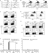
In vivo maturation of DCs and cross-presentation of α-GalCer in DCs after administration of tumor/Gal. (A) Spleen cells were obtained at 0, 2, 4, and 6 h after administration of CD1dhi-B16/Gal. Low density spleen cells were stained with anti-CD11c–PE and CD8α-FITC (to identify DCs and their CD8+ or CD8 subsets) and with biotinylated isotype control or CD86 mAb, followed by streptavidin-allophycocyanin. (B) As in A, but DCs were tested in three different organs for CD86 expression 6 h after injecting the tumor cells indicated on the left. All data in A, B, and D are representative of two independent experiments (n = 2 per group). (C) Splenic DCs were analyzed by intracellular cytokine staining for IL-12p40 4 h after immunization with tumor/Gal, followed by 4 h of culture in brefeldin A. DCs were identified with CD11c-allophycocyanin and CD8α-FITC and were subsequently fixed and stained with PE-conjugated anti–IL-12p40 mAb. (D and E) DC maturation studies, as in A and C, were performed in WT and Jα18−/− mice to show the NKT cell dependence. Numbers in C and E indicate the percentage of IL-12–producing cells in CD8+ or CD8 subsets of CD11c+ DCs, and all data are representative of three independent experiments (n = 3 per group). (F) To evaluate the cross-presentation of α-GalCer derived from tumor/Gal, DCs or non-DCs were isolated by CD11c magnetic beads at 10 h after injecting 5 × 10⁵ CD1dhi-B16/Gal into WT C57BL/6 or CD1d−/− mice. 105 APCs per well were co-cultured with 105 liver MNCs per well from C57BL/6 or Jα18−/− mice for 48 h. Then, the IFN-γ from the supernatants was measured by ELISA. All data are means ± SEM obtained from three independent experiments (n = 5 per group). *, P < 0.05 for WT versus Jα18−/− for liver MNCs; **, P < 0.01 for WT versus CD1d−/− for APCs.
Figure 7.
Figure 7.
DCs are required for inducing immunity to tumor/Gal. (A) 10 h after injecting CD1dhi-B16/Gal tumor cells, CD11c+ and CD11c cells were isolated, and 3 × 10⁶ CD11c+ and 5 × 10⁶ CD11c cells were transferred to naive C57BL/6 mice; spleen cells from uninjected mice served as the negative control. 1 wk later, we measured CD8+ T cells specific for melanoma antigens (trp2 and gp100) by ELISPOT assay. (B) 1 d after injecting CD1dhi-B16/Gal i.v. into CD11c-DTR transgenic mice, CD11c+ DCs were depleted with DT. 7 d later, we analyzed antigen-specific T cell responses to trp2 or gp100 peptides by ELISPOT assays. All data are means obtained from two independent experiments (n = 5 per group). *, P < 0.05 for CD11c+ transferred mice versus others (left) and DT-treated CD11c-DTR mice versus immunized WT mice (right). SFC, spot-forming cells.

References

    1. Blattman, J.N., and P.D. Greenberg. 2004. Cancer immunotherapy: a treatment for the masses. Science. 305:200–205. - PubMed
    1. Yee, C., J.A. Thompson, D. Byrd, S.R. Riddell, P. Roche, E. Celis, and P.D. Greenberg. 2002. Adoptive T cell therapy using antigen-specific CD8+ T cell clones for the treatment of patients with metastatic melanoma: in vivo persistence, migration, and antitumor effect of transferred T cells. Proc. Natl. Acad. Sci. USA. 99:16168–16173. - PMC - PubMed
    1. Dudley, M.E., J.R. Wunderlich, P.F. Robbins, J.C. Yang, P. Hwu, D.J. Schwartzentruber, S.L. Topalian, R. Sherry, N.P. Restifo, A.M. Hubicki, et al. 2002. Cancer regression and autoimmunity in patients after clonal repopulation with antitumor lymphocytes. Science. 298:850–854. - PMC - PubMed
    1. Morgan, R.A., M.E. Dudley, J.R. Wunderlich, M.S. Hughes, J.C. Yang, R.M. Sherry, R.E. Royal, S.L. Topalian, U.S. Kammula, N.P. Restifo, et al. 2006. Cancer regression in patients after transfer of genetically engineered lymphocytes. Science. 314:126–129. - PMC - PubMed
    1. Casares, N., M.O. Pequignot, A. Tesniere, F. Ghiringhelli, S. Roux, N. Chaput, E. Schmitt, A. Hamai, S. Hervas-Stubbs, M. Obeid, et al. 2005. Caspase-dependent immunogenicity of doxorubicin-induced tumor cell death. J. Exp. Med. 202:1691–1701. - VSports手机版 - PMC - PubMed

Publication types