"V体育官网入口" Skip to main page content
U.S. flag

An official website of the United States government

Dot gov

The VSports app下载. gov means it’s official. Federal government websites often end in . gov or . mil. Before sharing sensitive information, make sure you’re on a federal government site. .

Https

The site is secure. The https:// ensures that you are connecting to the official website and that any information you provide is encrypted and transmitted securely V体育官网. .

. 2008 Jan 15;111(2):723-31.
doi: 10.1182/blood-2007-05-091173. Epub 2007 Oct 1.

"V体育ios版" Calmodulin-dependent kinase IV links Toll-like receptor 4 signaling with survival pathway of activated dendritic cells

Affiliations

Calmodulin-dependent kinase IV links Toll-like receptor 4 signaling with survival pathway of activated dendritic cells

"VSports app下载" Maddalena Illario et al. Blood. .

Abstract

Microbial products, including lipopolysaccharide (LPS), an agonist of Toll-like receptor 4 (TLR4), regulate the lifespan of dendritic cells (DCs) by largely undefined mechanisms VSports手机版. Here, we identify a role for calcium-calmodulin-dependent kinase IV (CaMKIV) in this survival program. The pharmacologic inhibition of CaMKs as well as ectopic expression of kinase-inactive CaMKIV decrease the viability of monocyte-derived DCs exposed to bacterial LPS. The defect in TLR4 signaling includes a failure to accumulate the phosphorylated form of the cAMP response element-binding protein (pCREB), Bcl-2, and Bcl-xL. CaMKIV null mice have a decreased number of DCs in lymphoid tissues and fail to accumulate mature DCs in spleen on in vivo exposure to LPS. Although isolated Camk4-/- DCs are able to acquire the phenotype typical of mature cells and release normal amounts of cytokines in response to LPS, they fail to accumulate pCREB, Bcl-2, and Bcl-xL and therefore do not survive. The transgenic expression of Bcl-2 in CaMKIV null mice results in full recovery of DC survival in response to LPS. These results reveal a novel link between TLR4 and a calcium-dependent signaling cascade comprising CaMKIV-CREB-Bcl-2 that is essential for DC survival. .


"VSports" Figures
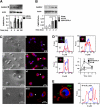
Figure 1
Figure 1
CaMKIV accumulates during differentiation of monocyte-derived dendritic cells. (A) CD14+ mononuclear cells were cultured in the presence of GM-CSF and IL-4. Whole-cell lysates were prepared at the indicated times and analyzed by immunoblot with specific antibodies (CaMKIV and actin). Aliquots of cells were used to measure CaMKIV and actin mRNA levels by quantitative reverse transcription–polymerase chain reaction. Bottom panel shows mean (± SD) of optical density measurements expressed as the ratio between the CaMKs and actin bands (n = 4). *P < .01. (B) Calpain regulates CaMKIV accumulation in differentiating monocytes. CD14+ mononuclear cells were cultured in the presence of ALLM, a selective calpain inhibitor (ALLM), or GM-CSF and IL-4 and analyzed for CaMKIV expression by immunoblot (top). The bottom panel shows mean (± SD) of the optical density measurements expressed as the ratio between the CaMKIV and actin bands (n = 4). *P < .01. (C-E) Intracellular distribution of CaMKIV in differentiating monocytes. (C) Transmission and confocal fluorescent immunocytochemistry images of CaMKIV expression in monocytes cultured for: 2 hours in regular medium (i,ii); 2 hours in the presence of GM-CSF/IL-4 or ALLM (iii, iv, v, and vi, respectively); and 120 hours in the presence of GM-CSF/IL-4 (vii,viii). (D) Line profiles of cells indicated by white arrows in the corresponding subpanels in panel C. The line segment is 20 μM; F indicates the fluorescence intensity in arbitrary units (a.u.). The bottom right graph shows the ratio (R) between the mean fluorescence intensity of Alexa Fluor 594 (CaMKIV) and Hoechst 33342 (nuclear staining) in the nuclear region along different line profiles. Means (± SD) represent 20 independent line profiles. *P < .01. (E) Expression and line profile analysis of CaMKIV in monocytes treated for 120 hours with GM-CSF/IL-4..
Figure 2
Figure 2
CaMKs regulate terminal differentiation and survival of monocyte-derived dendritic cells. Immature monocyte–derived DCs were cultured untreated or stimulated with LPS- (1 μg/μL) in the presence or absence of KN93 (10 μM), a selective inhibitor of the multifunctional CaMKs. After 24 hours, cells were recovered and double-stained with anti-CD86/anti-CD83 antibodies or with Annexin V/propidium iodide (A,B top). (A) Bottom: effects of KN93 on CD83 and CD86 expression as a function of the LPS dose. Mean (± SD) represents 6 independent experiments. (B) Bottom: effects of KN93 on survival of LPS-stimulated DC (LPS, 10 μg/mL) as a function of time or KN93 dose (left or right, respectively). Viability was calculated by trypan blue exclusion. Mean (± SD) represents 6 independent experiments.*P < .01. Values in panel A represent the mean fluorescente intensity of CD86 and the percentage of CD83 positive cells. Ctr refers to profiles of unstained cells. Values in panel B represent the percentage of cells in each quadrant.
Figure 3
Figure 3
CaMKIV regulates survival of monocyte–derived DCs. Monocyte-derived DCs were infected with Lenti-IRES-GFP lentivirus expressing Camk4, Camk4-WT, or Camk4-K71M (Mock, WT, and DN, respectively). After 48 hours, cells were cultured for an additional 18 hours in the presence of LPS (1 μg/mL) or left untreated. Top: fluorescence-activated cell sorting (FACS) profiles of DC stained with CD86, CD83, and Annexin-V.
Figure 4
Figure 4
The number of splenic mature DCs is reduced in Camk4−/− mice. Splenocytes from normal or Camk4−/− mice were counted and stained for CD11c and CD8. (A) Left panel: immunoblots show CaMKIV and actin expression in splenocytes isolated from 2 mice from each genotype. Middle: bar graph reports mean (± SD) of the total number of splenocytes (n = 15 mice per genotype). Right: percentage of WT and Camk4−/− CD11c + subsets. Bars graphs show mean (± SD) representing 15 mice per genotype. *P < .01. (B) LPS-induced mature DC accumulation is impaired in Camk4−/− mice in vivo. LPS or phosphate-buffered saline (PBS) was injected into Camk4−/− and control WT mice. Eighteen hours later, splenocytes were isolated and triple-stained with anti-CD11b, -CD11c and -I-A antibodies. For the typical dot plot profiles, the inset values show the percentage of cells in the R6/R7 gates (CD11bhigh/CD11chigh and CD11blow/CD11chigh, respectively. FACS profile histograms show I-A expression. Inset values refer to the percentage of I-Ahigh cells in the R6/R7 gates. Values in parenthesis display the percentage of CD11bhigh/CD11chigh/I-Ahigh and CD11blow/CD11chigh/I-Ahigh in whole splenocytes. *P < .01.
Figure 5
Figure 5
CaMKIV is not required for terminal differentiation and cytokine synthesis induced by LPS. Isolated CD11c+ splenic DCs from WT and Camk4−/− were exposed to LPS (10 μg/mL) or left untreated (none). After 16 hours, cells were recovered and double-stained for I-A and CD86 (A). Aliquots of cells were stained for the presence of intracellular TNF-α and IL-6 (A,B).
Figure 6
Figure 6
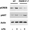
CaMKIV is required to link TLR4 signaling with pCREB accumulation. CD11c + DCs were isolated from spleens of WT or Camk4−/− mice and cultured in the presence or absence of LPS (10 μg/mL) for 1 hour. Whole lysates were separated by SDS-PAGE and immunoblotted with the reported antibodies (pCREB, pAKT, actin, LPS). A typical immunoblot analysis is shown.
Figure 7
Figure 7
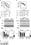
CaMKIV regulates lifespan and Bcl-2 family protein accumulation. CD11c + DCs were positively selected from WT (Camk4+/+), Camk4−/−, BCL-2tg/tg transgenic, and Camk4−/−/BCL-2tg/tg hybrid mice cultured in the presence or absence of LPS (10 μg/mL). (A,D) Viability was assayed by trypan blue exclusion at daily intervals. The results represent mean and SD of 6 independent experiments. (B,E) Typical results obtained by immunoblot analysis. Bar graphs show mean (± SD) of the optical density measurements expressed as the ratio between Bcl-2 or Bcl-xL and actin bands (n = 6). (C) Immunoblot shows the typical expression of CaMKIV and hu-Bcl-2 detected in WT (Camk4+/+), Camk4−/−, BCL-2tg/tg transgenic, and Camk4−/−/BCL-2tg/tg hybrid mice (lanes 1, 2, 3, and 4, respectively). *P < .01.

References

    1. Banchereau J, Steinman RM. Dendritic cells and the control of immunity. Nature. 1998;392:245–252. - PubMed
    1. Steinman RM. The dendritic cell system and its role in immunogenicity. Annu Rev Immunol. 1991;9:271–296. - PubMed
    1. Steinman RM, Bonifaz L, Fujii S, et al. The innate functions of dendritic cells in peripheral lymphoid tissues. Adv Exp Med Biol. 2005;560:83–97. - PubMed
    1. Akira S, Takeda K. Toll-like receptor signalling. Nat Rev Immunol. 2004;4:499–511. - PubMed
    1. De Smedt T, Pajak B, Muraille E, et al. Regulation of dendritic cell numbers and maturation by lipopolysaccharide in vivo. J Exp Med. 1996;184:1413–1424. - PMC - PubMed

Publication types

MeSH terms

Substances