Skip to main page content
U.S. flag

An official website of the United States government

Dot gov

The VSports app下载. gov means it’s official. Federal government websites often end in . gov or . mil. Before sharing sensitive information, make sure you’re on a federal government site. .

Https

The site is secure. The https:// ensures that you are connecting to the official website and that any information you provide is encrypted and transmitted securely. V体育官网.

. 2007 Sep 28:8:85.
doi: 10.1186/1471-2199-8-85.

Reciprocal regulation of p63 by C/EBP delta in human keratinocytes

Affiliations

V体育平台登录 - Reciprocal regulation of p63 by C/EBP delta in human keratinocytes

Serena Borrelli et al. BMC Mol Biol. .

Abstract (V体育官网入口)

Background: Genetic experiments have clarified that p63 is a key transcription factor governing the establishment and maintenance of multilayered epithelia. Key to our understanding of p63 strategy is the identification of target genes VSports手机版. We perfomed an RNAi screening in keratinocytes for p63, followed by profiling analysis. .

Results: C/EBPdelta, member of a family with known roles in differentiation pathways, emerged as a gene repressed by p63. We validated C/EBPdelta as a primary target of DeltaNp63alpha by RT-PCR and ChIP location analysis in HaCaT and primary cells. C/EBPdelta is differentially expressed in stratification of human skin and it is up-regulated upon differentiation of HaCaT and primary keratinocytes V体育安卓版. It is bound to and activates the DeltaNp63 promoter. Overexpression of C/EBPdelta leads to alteration in the normal profile of p63 isoforms, with the emergence of DeltaNp63beta and gamma, and of the TA isoforms, with different kinetics. In addition, there are changes in the expression of most p63 targets. Inactivation of C/EBPdelta leads to gene expression modifications, in part due to the concomitant repression of DeltaNp63alpha. Finally, C/EBPdelta is found on the p63 targets in vivo by ChIP analysis, indicating that coregulation is direct. .

Conclusion: Our data highlight a coherent cross-talk between these two transcription factors in keratinocytes and a large sharing of common transcriptional targets V体育ios版. .


Figures
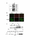
Figure 1
Figure 1
Validation of C/EBPδ as a target of p63. A. Evaluation of C/EBPα, C/EBPβ and C/EBPδ, by semi-quantitative RT-PCR analysis in control and cells treated with siRNA of p63. β-actin was used as an internal control. In the lower Panel, primary human keratinocytes (KCs) were treated similarly and RT-PCR performed for ΔNp63α and C/EBPδ; the invariant GAPDH was included as internal control. B. Confocal microscopy immunofluorescence analysis of primary KCs treated with control and p63 RNAi oligos. Fixation and staining with the indicated antibodies was performed at 48 hours post-transfections. Note that the laser settings of the C/EBPδ image of p63-inactivated cells had to be lowered considerably due to extemely strong fluorescence. C. Chromatin Immunoprecipitation analysis of the C/EBPδ-1 Kb (Indicated by the square box), using HaCaT cells (Left Panel) and the indicated antibodies: NF-YB, p63 (Santa Cruz H137; Dako 4A4) and control -Ctl- anti-Flag antibodies (Sigma). Right Panel, ChIPs with primary KCs, anti-p63 4A4 (Dako), anti-p63α specific polyclonal [19], anti-p53 (Ab7 Calbiochem). As a control, ChIPs from KCs were used to amplify the α-globin promoter. In the bottom part, the p53 and p63 sites at -1000 of the human C/EBPδ promoter are shown, in an area of conservation with the mouse gene. HBA1 is the human α-globin promoter.
Figure 2
Figure 2
Regulation of C/EBPδ in HaCaT and primary keratinocytes. A. Upper Panel. RT-PCR analysis of the C/EBPs in HaCaT cells before and after 3 and 72 hours in differentiation medium. cK1 was used as control for differentiation, NF-YB as internal control. Lower Panel. Western blot analysis of HaCat extracts with the indicated antibodies. B. Immunofluorescence analysis of growing and differentiated HaCaT [72 hours] using the indicated antibodies. cK1 was used to control for differentiation. Ctl refers to cells stained with the rabbit secondary antibody only. C. RT-PCR analysis as in A, except that primary human keratinocytes were used, before or after 3 days in differentiation medium. cK1 and cK14 were used as markers of differentiation. D. Same confocal experiments as in B, using human primary keratinocytes.
Figure 3
Figure 3
Regulation of C/EBPδ in human skin. Fixed samples of two representative human interfollicular skin sections were double stained with anti-p63 4A4 and the C/EBPδ antibodies. Analysis was performed by Immunofluorescence Confocal microscopy. Arrows in the merge Panel indicate nuclear staining of cells that are positive for both antibodies, in the basal layer. Yellow signals in the spinous layer refer to cells co-expressing the two factors. B, basal layer; S, spinous layer; G, granulous layer; C, corneum stratum. In the CTL Panels, we stained a skin section only with mouse and rabbit secondary antibodies used above.
Figure 4
Figure 4
C/EBPδ is bound to and regulates the ΔNp63 promoter. A. Left Panel. Human KCs were transfected with mouse C/EBPδ. RNAs were extracted at the indicated time and RT-PCR is shown for the indicated genes: controls (mC/EBPδ, GAPDH) and different p63 isoform (upper Panels). Right Panel. RNAi of C/EBPδ with three different oligos. C/EBPδ mRNA was checked for inhibition. B. Bioinformatic analsysis of the ΔNp63 promoters for C/EBP sites with different algorithms: a cluster of sites commonly detected at position -2400 is shown. Analysis was performed with rVista [63], CONSITE [64] and CUSTOM. Shown are also mouse conservation and a schematic representation of the amplified region. C. Transfection analysis of ΔNp63 constructs [22] without and with NF-Y and/or C/EBPδ. D. ChIP analysis of the upstream regions of ΔNp63 with the indicated antibodies, using chromatin from primary KCs.
Figure 5
Figure 5
Overexpression and functional inactivation of C/EBPδ in primary KCs. Human KCs were transfected with mouse C/EBPδ. RNAs were extracted at the indicated time and RT-PCR is shown for the indicated p63 target genes (Left Panels). In the Right Panels, C/EBPδ inactivation was performed in human KCs with siRNA oligonucleotide 2 of Fig. 4 and with a scramble oligo, in parallel. RT-PCR analysis of the genes in Left Panels is shown. The increase of c-Jun in RNAi-treated cells can only be visualized by low cycles RT-PCR, where a signal from the control oligo RNA is not detectable. On the Right part of the Figure, the effect of RNAi inactivation of p63 is depicted, according to published data and to Ref. 19.
Figure 6
Figure 6
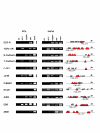
C/EBPδ binds to p63 targets. ChIP analysis of NF-Y, C/EBPδ and p63 on different genes targeted by p63 in HaCat cells (Right Panels) and in human primary KCs (Left Panels). The regions considered were described for p63 [19] and are detailed in the cartoon. The analysis of C/EBP (Triangles) consensus sequences according to the three algorithms used in Fig. 4 is depicted. Circles refer to the p63 consensus recently described by Orrt et al [65]. Slashed circles refer to p53 consensus. The position of the amplicons is indicated.
Figure 7
Figure 7
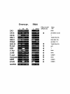
Regulation of C/EBPδ targets during differentiation. The targets of C/EBPδ are evaluated in primary KC before and after 72 hours of differentiation by semi-quantitative RT-PCR analysis.

References

    1. McKeon F. p63 and the epithelial stem cell: more than status quo? Genes Dev. 2004;18:465–469. doi: 10.1101/gad.1190504. - DOI - PubMed
    1. Mills AA, Zheng B, Wang X-J, Vogel H, Roop DR, Bradley A. p63 is a homologue required for limb and epidermal morphogenesis. Nature. 1999;398:708–713. doi: 10.1038/19531. - "V体育ios版" DOI - PubMed
    1. Yang A, Schweitzer R, Sun DQ, Kaghad M, Walker N, Bronson RT, Tabin C, Sharpe A, Caput D, Crum C, McKeon F. p63 is essential for regenerative proliferation in limb, craniofacial and epithelial development. Nature. 1999;398:714–718. doi: 10.1038/19539. - DOI - PubMed
    1. Bakkers J, Hild M, Kramer C, Furutani-Seiki M, Hammerschmidt M. Zebrafish DeltaNp63 is a direct target of Bmp signaling and encodes a transcriptional repressor blocking neural specification in the ventral ectoderm. Dev Cell. 2002;2:617–627. doi: 10.1016/S1534-5807(02)00163-6. - DOI - PubMed
    1. Rinne T, Hamel B, Bokhoven HV, Brunner HG. Pattern of p63 mutations and their phenotypes-update. Am J Med Genet A. 2006;140:1396–406. - PubMed

Publication types

MeSH terms (VSports app下载)

VSports - Substances