VSports app下载 - Skip to main page content
U.S. flag

An official website of the United States government

Dot gov

The . gov means it’s official. Federal government websites often end in . gov or . mil. Before sharing sensitive information, make sure you’re on a federal government site VSports app下载. .

Https

The site is secure V体育官网. The https:// ensures that you are connecting to the official website and that any information you provide is encrypted and transmitted securely. .

. 2007 Sep 21;130(6):1005-18.
doi: 10.1016/j.cell.2007.07.020.

Heat shock factor 1 is a powerful multifaceted modifier of carcinogenesis

Affiliations

Heat shock factor 1 is a powerful multifaceted modifier of carcinogenesis

Chengkai Dai et al. Cell. .

"V体育ios版" Abstract

Heat shock factor 1 (HSF1) is the master regulator of the heat shock response in eukaryotes, a very highly conserved protective mechanism. HSF1 function increases survival under a great many pathophysiological conditions. How it might be involved in malignancy remains largely unexplored. We report that eliminating HSF1 protects mice from tumors induced by mutations of the RAS oncogene or a hot spot mutation in the tumor suppressor p53. In cell culture, HSF1 supports malignant transformation by orchestrating a network of core cellular functions including proliferation, survival, protein synthesis, and glucose metabolism. The striking effects of HSF1 on oncogenic transformation are not limited to mouse systems or tumor initiation; human cancer lines of diverse origins show much greater dependence on HSF1 function to maintain proliferation and survival than their nontransformed counterparts. While it enhances organismal survival and longevity under most circumstances, HSF1 has the opposite effect in supporting the lethal phenomenon of cancer. VSports手机版.


Figures

Figure 1
Figure 1. HSF1 Deficiency Suppresses Chemical Skin Carcinogenesis
(A) Representative images of mouse skin tumors 25 weeks after topical DMBA application. (B) Lower skin tumor incidence and longer incubation time in Hsf1-/- mice (p < 0.0001, two-way ANOVA). (C) Lower tumor burden in Hsf1-/- mice. The data are presented as the number of skin tumors per mouse (mean ± SE, p < 0.0001, two-way ANOVA). (D) Smaller tumor volumes in Hsf1-/- mice (the lines indicate geometric means; p = 0.0003, Mann Whitney test). (E) The survival curves of Hsf1+/+ and Hsf1-/- mice following skin carcinogenesis (median survival: Hsf1+/+ 41 weeks; Hsf1-/- undefined; p = 0.0073, Logrank test).
Figure 2
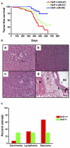
Figure 2. HSF1 Deficiency Suppresses Tumorigenesis Driven by Mutant p53
(A) Tumor-free survival curves of p53R172H knockin mice (Hsf1+/+ versus Hsf1-/-, p = 0.0001; Hsf1+/- versus Hsf1-/-, p = 0.0185;Hsf1+/+ versus Hsf1+/- , p = 0.0387; Logrank test). (B) Representative micrographs of tumors (scale bars, 160 mm). (Ba) Carcinoma, (Bb) lymphoma, (Bc) soft tissue sarcoma, (Bd) teratoma (A, adipose; B, bone; M, muscle; RE, respiratory epithelium). (C) Tumor spectra of Hsf1+/+ and Hsf1+/- mice(Hsf1+/+, n = 15; Hsf1+/- , n = 10).
Figure 3
Figure 3. HSF1 Enables Cellular Transformation Initiated by Oncogenic RAS and PDGF-B
(A) Hsf1-/- MEFs are relatively resistant to focus formation driven by oncogenic H-RASV12D. Immortalized MEFs were plated and transduced with retroviruses encoding the genes indicated. Foci were fixed and visualized by dye staining. The number of foci per well was quantified as shown in Figure S2. All experiments were repeated once with similar results. (B) Hsf1-/- MEFs are relatively resistant to focus formation driven by the proto-oncogene PDGF-B. (C) Hsf1-/- MEFs are refractory to proliferation driven by oncogenic RAS and PDGF-B. Equal numbers of immortalized Hsf1+/+ and Hsf1-/- MEFs were transduced with retroviruses encoding GFP, H-RASV12D,or PDGF-B. The cells were fixed on the indicated days and the number of cells per well determined by fluorescent DNA staining. Relative cell number was calculated by normalizing the values against the GFP-transduced group at each time point (mean ± SD, n = 5, ***p < 0.001, two-way ANOVA). (D) Expression of c-MYC and LTA does not drive marked proliferation in immortalized Hsf1+/+ and Hsf1-/- MEFs. (E) Hsf1-/- MEFs show no enhanced survival in response to RAS and PDGF/B expression but reduced survival in response to c-MYC and LTA expression. Viability of immortalized Hsf1+/+ and Hsf1-/- MEFs was determined by flow cytometry 36 hr after transduction. The data are presented as percent nonviable cells (mean ± SD, n = 5, *p < 0.05; ***p < 0.001, Student’s t test).
Figure 4
Figure 4. HSF1 Modulates Signal Transduction
(A) Hsf1-/- and C2-transduced MEFs show remarkably decreased expression of KSR1 protein by immunoblotting. Ponceau red staining was used to confirm equal protein loading. (B) Hsf1-/- MEFs display blunted ERK activation in response to serum stimulation. Cells were stimulated with 20% FBS for the periods of time indicated, then fixed and stained with phospho-ERK1/2 antibody (green) and the DNA stain TO-PRO-3 iodide (red), which was used to normalize for relative cell number. The plate was scanned, and data are presented as relative phospho-ERK1/2 level by setting values at 0 min as 100% (mean ± SD, n = 5, **p < 0.01; ***p < 0.001; two-way ANOVA). (C) Hsf1-/- and Hsf1 knockdown cells show markedly reduced phosphorylation of endogenous PKA substrates. Equal amounts of total protein were loaded and probed with antibodies recognizing phospho-(Ser/Thr) PKA substrate, HSF1, and GAPDH, respectively. (D) Hsf1-/- MEFs possess lower PKA activity. The PKA activity in lysates prepared from Hsf1+/+ and Hsf1-/- MEFs was measured using the classical substrate Kemptide. Reaction mixtures were fractionated by agarose gel electrophoresis to visualize the extent of substrate phosphorylation. PKA, recombinant PKA as positive control. NC, peptide substrate only without lysate or recombinant PKA. The relative amount of phosphorylated peptide in each lane was quantitated by fluorometry.
Figure 5
Figure 5. HSF1 Modulates Translation Machinery
(A and B) HSF1 maintains p70 S6K phosphorylation and ribosomal protein expression during serum starvation. Cells were first serum starved for 2 days, then either stimulated with 20% FBS overnight or maintained under serum-free conditions. (C) Hsf1-/- MEFs are more sensitive to the proliferation-inhibitory effect of rapamycin. Cells were exposed to a series of rapamycin concentrations as indicated for 5 days. After fixation, the relative number of cells in each well was determined by DNA staining. Data are presented as percent control by normalizing the values of rapamycin-treated wells against those of solvent-treated wells (mean ± SD, n = 5, **p < 0.01, two-way ANOVA). (D) Hsf1-/- MEFs are more susceptible to cell-cycle arrest induced by rapamycin. Cells were treated with either methanol vehicle or rapamycin (100 nM) for 24 hr. Cell-cycle distribution was determined by flow cytometry. Data are presented as percent cells in each phase of the cell cycle (mean ± SD, n = 3, ***p < 0.001, two-way ANOVA).
Figure 6
Figure 6. HSF1 Modulates Glucose Metabolism
(A and B) HSF1 regulates glucose uptake. Cells were incubated in medium containing either PBS (dotted lines) or 100 μM 2-NBDG (solid lines) overnight. The extent of glucose uptake was measured by flow cytometry. Data are presented as frequency histograms of relative tracer uptake. (A) depicts Hsf1+/+ (red line) and Hsf1-/- MEFs (blue line). (B) depicts C2 (blue line)- and C3 (red line)-transduced MEFs. The mean fluorescence intensity of each cell population is indicated (mean ± SD, n = 5, Hsf1+/+ versus Hsf1-/- 2-NBDG p < 0.0001; C3 versus C2 2-NBDG p < 0.0001, Student’s t test). (C and D) Hsf1-/- MEFs are relatively resistant to glucose deprivation. Cells were incubated with either glucose-replete or -reduced medium for 4 days. The number and viability of cells in each well were determined by flow cytometry. Data are presented as relative cell numbers and percent nonviable cells (mean ± SD, n = 5, ***p < 0.001, two-way ANOVA).
Figure 7
Figure 7. HSF1 Is Required for Maintenance of the Transformed Phenotype
(A) ShRNA constructs exhibit differential HSF1-targeting efficacy. 293T cells were transfected with equal amounts of the indicated shRNA plasmid DNAs, and MCF-7 cells were transduced with the same shRNA constructs but packaged as lentiviral supernatants. Cells were harvested for immunoblotting after 3 days of puromycin selection. (B) The effect of HSF1 compromise on cell growth and survival correlates with malignant state. Cells were plated in 96-well format and transduced with viral supernatants as indicated. Viable cell number in each well was measured 4 days after viral transduction. Data are presented as relative viable cell number by normalizing the values of each transduction group against those of GFP shRNA-transduced wells (mean ± SD, n = 5, *p < 0.05, **p < 0.01, ***p < 0.001, two-way ANOVA). (C) Compromise of HSF1 impairs the growth and survival of established human breast cancer cells (mean ± SD, n = 5, *p < 0.05, **p < 0.01, ***p < 0.001, two-way ANOVA). (D) Compromise of HSF1 impairs the growth and survival of human cancer cells derived from diverse histological origins (mean ± SD, n = 5, *p < 0.05, **p < 0.01, ***p < 0.001, two-way ANOVA). All experiments were repeated at least once with similar results.

Comment in

References

    1. Auluck PK, Meulener MC, Bonini N. Mechanisms of suppression of {alpha}-synuclein neurotoxicity by geldanamycin in Drosophila. J. Biol. Chem. 2005;280:2873–2878. - "VSports最新版本" PubMed
    1. Balmain A, Ramsden M, Bowden GT, Smith J. Activation of the mouse cellular Harvey-ras gene in chemically induced benign skin papillomas. Nature. 1984;307:658–660. - PubMed (V体育平台登录)
    1. Beausejour CM, Campisi J. Ageing: balancing regeneration and cancer. Nature. 2006;443:404–405. - PubMed
    1. Birch-Machin I, Gao S, Huen D, McGirr R, White RA, Russell S. Genomic analysis of heat-shock factor targets in Drosophila. Genome Biol. 2005;6:R63. Published online June 10, 2005. 10.1186/gb-2005-6-7-r63. - PMC - PubMed
    1. Bissell MJ, Rambeck WA, White RC, Bassham JA. Glycerol phosphate shuttle in virus-transformed cells in culture. Science. 1976;191:856–858. - VSports最新版本 - PubMed

Publication types

MeSH terms