Skip to main page content (V体育安卓版)
U.S. flag

An official website of the United States government

Dot gov

The . gov means it’s official. Federal government websites often end in . gov or . mil. Before sharing sensitive information, make sure you’re on a federal government site VSports app下载. .

Https

The site is secure. The https:// ensures that you are connecting to the official website and that any information you provide is encrypted and transmitted securely V体育官网. .

. 2007 Aug;15(8):893-903.
doi: 10.1016/j.str.2007.06.015.

An IgG-like domain in the minor pilin GBS52 of Streptococcus agalactiae mediates lung epithelial cell adhesion

Affiliations

VSports app下载 - An IgG-like domain in the minor pilin GBS52 of Streptococcus agalactiae mediates lung epithelial cell adhesion

Vengadesan Krishnan et al. Structure. 2007 Aug.

Abstract

Streptococcus agalactiae is the leading cause of neonatal pneumonia, sepsis, and meningitis VSports手机版. The pathogen assembles heterotrimeric pilus structures on its surface; however, their function in pathogenesis is poorly understood. We report here the crystal structure of the pilin GBS52, which reveals two IgG-like fold domains, N1 and N2. Each domain is comprised of seven antiparallel beta strands, an arrangement similar to the fold observed in the Staphylococcus aureus adhesin Cna. Consistent with its role as an adhesin, deletion of gbs52 gene significantly reduces bacterial adherence to pulmonary epithelial cells. Moreover, latex beads linked to the GBS52 protein adhere to pulmonary but not to many other epithelial cells; binding to the former is specifically inhibited by antibodies against GBS52. Nonetheless, substantial binding is only observed with N2 domain-conjugated beads. This study presents the structure of a Gram-positive pilin that utilizes a distinct IgG fold variant to mediate pathogen adherence to a specific tissue. .


Figures

Figure 1
Figure 1. Minor Pilins Are Required for Streptococcal Adherence
(A) Graphic representation of GBS pilus island 1 (PI-1) from strain 2603V/R with three sortase genes, along with three sortase-mediated pilus assembly genes (gbs80, gbs52, and gbs104). (B) Requirement of GBS52 for streptococcal adhesion to lung epithelial cells. Confluent monolayers of lung epithelial cells (A549) were infected with the wild-type and its isogenic deletion mutants, and adherent bacteria were then enumerated. Data are presented as percentage of adhesion relative to that of wild-type. The results are presented as averages (with standard deviations; ±SD) from at least three independent experiments performed in quadruplicate.
Figure 2
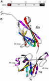
Figure 2. The Structure of the Minor Pilin GBS52
(A) Schematic domain organization of GBS52. GBS52 contains an N-terminal signal sequence (SS), followed by the 120 residue N1 domain, 116 residue N2 domain, and a C-terminal cell wall sorting signal (CWS). (B) Ribbon diagram of the overall crystal structure of GBS52. The two domains N1 and N2 are placed vertically to the plane of the paper. The strands of both N1 and N2 domains are assigned with rainbow colors (from red [A] to violet [G]) for comparison with other topologically related domains.
Figure 3
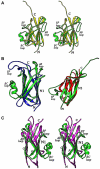
Figure 3. GBS52 Domain Comparison
(A) A stereo-view ribbon diagram of the superposition of the N1 domain (green) and the N2 domain (yellow) of GBS52. The variable regions are labeled (AB, BC, and EF loops and C-terminal tail). (B) Comparison of GBS52 domains with other best topologically and structurally related domains in the Protein Data Bank as found by the Dali server (Dietmann et al., 2001). Left: superposition of the N1 domain (green) of GBS52 and first b sandwich domain (SD1) of Ferovidolysin (in blue; PDB ID code 1R6V). Right: superposition of the N2 domain (green) of GBS52 and C-terminal subdomain of Carboxypeptidase D domain II (in red; PDB ID code 1QMU). (C) A stereo view of structural comparison of the N1 domain of GBS52 (green) with the Cna B region subdomain D1 (magenta). Main differences are seen in the loops and strand F position.
Figure 4
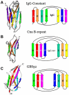
Figure 4. Topology Comparison
Arrows represent the β strands colored similarly to the Figure 2 representation. Shown are the IgG constant domain (A), the D1 domain of the B1 region of S. aureus collagen-binding protein Cna (B), and the N1 domain of GBS52 (C). The D1 resembles the IgG-like structure with a four- and three-β-stranded barrel, but exhibits a novel IgG-rev fold. The β sandwich structures are compared as follows: (IgG/D1) A/F, D/C, C/D, B/E, E/B, and F/A. The strands of IgG and D1 are aligned similarly to that presented in a previously published report (Deivanayagam et al., 2000). If any one of the colors on IgG and D1 are aligned, an adjacent strand of D1 (to the right or left) is observed to be on the opposite side in IgG (Deivanayagam et al., 2000). The topology of GBS52 is very similar to that of D1, except that the F strand in GBS52 is shifted to the opposite β sheet, resulting in DAG and CBEF stranded sheets instead of the DAGF and CBE stranded sheets observed in the D1 domain.
Figure 5
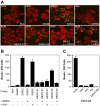
Figure 5. GBS52-Mediated Adherence Specifically to Human Lung Epithelial Cells, A549
(A) Semiconfluent cells grown on coverslips were treated with protein-coated beads. The washed cells were fixed and stained with Texas red-X phalloidin. Shown here are fluorescence images of cells incubated with fluorescent beads bound to BSA, GBS80, GBS52, or GBS52 with HeLa cells, the N1 domain of GBS52, the N2 domain of GBS52, or GBS52 blocked with α-GBS52, or GBS52 blocked with an unrelated antibody against a corynebacterial sortase, α-SrtA. (B) The results are presented as an average of the number of beads bound per 200 cells of three independent experiments (with standard deviations; ±SD). (C) Experiments performed as described in (A), except that indicated cells were treated with GBS52 protein-coated beads.

References

    1. Baker C, Edward M. Group B streptococcal infections. In: Remington JS, Klein JO, editors. Infectious Diseases of the Fetus and Newborn Infant. W.B. Saunders; Philadelphia: 2001. pp. 1091–1156.
    1. Barocchi MA, Ries J, Zogaj X, Hemsley C, Albiger B, Kanth A, Dahlberg S, Fernebro J, Moschioni M, Masignani V, et al. A pneumococcal pilus influences virulence and host inflammatory responses. Proc. Natl. Acad. Sci. USA. 2006;103:2857–2862. - V体育ios版 - PMC - PubMed
    1. Brunger AT, Adams PD, Clore GM, DeLano WL, Gros P, Grosse-Kunstleve RW, Jiang JS, Kuszewski J, Nilges M, Pannu NS, et al. Crystallography & NMR System: a new software suite for macromolecular structure determination. Acta Crystallogr. D Biol. Crystallogr. 1998;54:905–921. - PubMed
    1. CCP4 (Collaborative Computational Project, Number 4) The CCP4 suites: programs for protein crystallography. Acta Crystallogr. D Biol. Crystallogr. 1994;50:760–763. - PubMed
    1. Cieslewicz MJ, Kasper DL, Wang Y, Wessels MR. Functional analysis in type Ia group B Streptococcus of a cluster of genes involved in extracellular polysaccharide production by diverse species of streptococci. J. Biol. Chem. 2001;276:139–146. - PubMed

Publication types

MeSH terms

Associated data (VSports注册入口)

LinkOut - more resources