Skip to main page content
U.S. flag

An official website of the United States government

Dot gov

The VSports app下载. gov means it’s official. Federal government websites often end in . gov or . mil. Before sharing sensitive information, make sure you’re on a federal government site. .

Https

The site is secure V体育官网. The https:// ensures that you are connecting to the official website and that any information you provide is encrypted and transmitted securely. .

. 2007 Sep 21;372(3):764-73.
doi: 10.1016/j.jmb.2007.06.059. Epub 2007 Jun 29.

V体育ios版 - The structure of isolated Synechococcus strain WH8102 carboxysomes as revealed by electron cryotomography

Affiliations

The structure of isolated Synechococcus strain WH8102 carboxysomes as revealed by electron cryotomography (V体育ios版)

Cristina V Iancu et al. J Mol Biol. .

Abstract

Carboxysomes are organelle-like polyhedral bodies found in cyanobacteria and many chemoautotrophic bacteria that are thought to facilitate carbon fixation. Carboxysomes are bounded by a proteinaceous outer shell and filled with ribulose 1,5-bisphosphate carboxylase/oxygenase (RuBisCO), the first enzyme in the CO(2) fixation pathway, but exactly how they enhance carbon fixation is unclear. Here we report the three-dimensional structure of purified carboxysomes from Synechococcus species strain WH8102 as revealed by electron cryotomography. We found that while the sizes of individual carboxysomes in this organism varied from 114 nm to 137 nm, surprisingly, all were approximately icosahedral. There were on average approximately 250 RuBisCOs per carboxysome, organized into three to four concentric layers. Some models of carboxysome function depend on specific contacts between individual RuBisCOs and the shell, but no evidence of such contacts was found: no systematic patterns of connecting densities or RuBisCO positions against the shell's presumed hexagonal lattice could be discerned, and simulations showed that packing forces alone could account for the layered organization of RuBisCOs. VSports手机版.


Figures

Fig. 1
Fig. 1. Example image and reconstruction slice
(a) The nominally untilted image from a single-axis tilt series. (b) 6.7 nm-thick slice through the corresponding (undenoised) 3-D reconstruction. Scale bar 100 nm.
Fig. 2
Fig. 2. Carboxysomes are icosahedral
Four individual reconstructed carboxysomes are shown (one per row). From left to right appear the manually segmented surface, the best-fitting regular icosahedron, and three orthogonal central slices (xy, yz, and xz) through the denoised carboxysomes. The central slices are surrounded by the fit icosahedra, enlarged slightly and with the same color-coded faces to allow comparison of the actual shells with the corresponding cross sections of regular icosahedron. The anisotropic resolution is due to the limited tilt range accessible in ECT. Scale bar 50 nm.
Fig. 3
Fig. 3. Histogram of carboxysome diameters
Each bin is 1 nm wide. The diameter of icosahedra with all possible T-numbers in the same size range built from hexagons of edge length 4 nm (corresponding to crystal structure PDB 2AIB of CcmK2) are also shown.
Fig. 4
Fig. 4. Radial density profiles
Radial density profiles of individual carboxysomes (thin lines) and their average (thick line), scaled to normalize the densities of the outermost peaks (the shell). The positions of the well-defined peaks (the three internal RuBisCO layers and the carboxysome shell) are marked with arrows. For absolute scale, the average radius was 62 nm.
Fig. 5
Fig. 5. Identification of individual RuBisCOs through template-matching
Densities identified as RuBisCO by template matching followed by a customized peak search are circled in red on 6.7 nm slices through the (a) undenoised and (b) denoised carboxysome. (c) 3-D representation of the same carboxysome in which the densities have been replaced by the RuBisCO template.
Fig. 6
Fig. 6. Number of RuBisCOs per carboxysome as a function of carboxysome volume
The number of RuBisCOs was assessed by template matching followed by a customized peak-search algorithm, and the volume calculated as that of the best fitting regular icosahedron.
Fig. 7
Fig. 7. Relative arrangement of RuBisCOs and the presumed hexagonal lattice of the shell
All the densities (red) in the outermost layer of RuBisCO were projected on the faces of the icosahedron fit to the carboxysome shell. The presumed hexagonal lattice (with hexagonal spacing of 7 nm) for the corresponding T=81 icosahedron is shown in blue. No correspondence was discerned.
Fig. 8
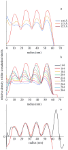
Fig. 8. Spontaneous generation of nested layers in icosahedral packing simulations
Icosahedral containers of average size (123 nm) were filled with randomly diffusing hard spheres of various diameters spanning the range of molecular dimensions present in RuBisCO. The resulting radial average density profiles from 14 independent packing simulations are shown in each case. (a) Profiles for 304 spheres of different diameters. The spheres spontaneously pack into layers in all cases. (b) Profiles for different numbers of 12.3 nm diameter spheres. As the number of spheres gets larger, the layers move slightly closer to the container boundary. (c) The average profile from the experimentally reconstructed carboxysomes (including an additional outer peak corresponding to the shell) (black) and the profile of the closest matching simulation, a 123 nm container filled with three hundred and four 12.3 nm spheres (red).

References

    1. Tcherkez GGB, Farquhar GD, Andrews TJ. Despite slow catalysis and confused substrate specificity, all ribulose bisphosphate carboxylases may be nearly perfectly optimized. Proc Nat Acad Sci USA. 2006;103:7246–7251. - PMC - PubMed
    1. Badger MR, Price GD, Long BM, Woodger FJ. The environmental plasticity and ecological genomics of the cyanobacterial CO2 concentrating mechanism. J Exp Bot. 2006;57:249–265. - PubMed
    1. Price GD, Sültemeyer D, Klughammer B, Ludwig M, Badger MR. The function of the CO2 concentrating mechanism in several cyanobacterial strains: a review of general physiological characteristics, genes, proteins, and recent advances. Can J Bot. 1998;76:973–1002.
    1. Heinhorst S, Cannon G, Shively JM. Carboxysomes and carboxysome-like inclusions. In: Shively JM, editor. Microbiology Monographs, Complex intracellular structures in prokaryotes. Vol. 2. Springer; Berlin, Heidelberg, New York: 2006. pp. 141–166.
    1. Badger MR, Hanson D, Price GD. Evolution and diversity of CO2 concentrating mechanisms in cyanobacteria. Funct Plant Biol. 2002;29:161–173. - PubMed

V体育安卓版 - Publication types

Substances