<kbd date-time="CQWqtQ6w"></kbd><del id="MaZO0cf"></del> Skip to main page content
U.S. flag

An official website of the United States government

Dot gov

The . gov means it’s official VSports app下载. Federal government websites often end in . gov or . mil. Before sharing sensitive information, make sure you’re on a federal government site. .

Https

The site is secure. The https:// ensures that you are connecting to the official website and that any information you provide is encrypted and transmitted securely. V体育官网.

. 2007 Sep;81(18):9748-58.
doi: 10.1128/JVI.01122-07. Epub 2007 Jul 3.

Manipulation of the toll-like receptor 7 signaling pathway by Epstein-Barr virus

Affiliations

V体育官网入口 - Manipulation of the toll-like receptor 7 signaling pathway by Epstein-Barr virus

Heather J Martin et al. J Virol. 2007 Sep.

"V体育安卓版" Abstract

Epstein-Barr virus (EBV) infection of primary B cells causes B-cell activation and proliferation. Activation of B cells requires binding of antigen to the B-cell receptor and a survival signal from ligand-bound CD40, signals that are provided by the EBV LMP1 and LMP2A latency proteins. Recently, Toll-like receptor (TLR) signaling has been reported to provide a third B-cell activation stimulus VSports手机版. The interaction between the EBV and TLR pathways was therefore investigated. Both UV-inactivated and untreated EBV upregulated the expression of TLR7 and downregulated the expression of TLR9 in naive B cells. UV-inactivated virus transiently stimulated naive B-cell proliferation in the presence of the TLR7 ligand R837, while addition of the TLR7 antagonist IRS 661 impaired cell growth induced by untreated EBV. Interferon regulatory factor 5 (IRF-5) is a downstream mediator of TLR7 signaling. IRF-5 was induced following EBV infection, and IRF-5 was expressed in B-cell lines with type III latency. Expression of IRF-5 in this setting is surprising since IRF-5 has tumor suppressor and antiviral properties. B-cell proliferation assays provided evidence that EBV modulates TLR7 signaling responses. Examination of IRF-5 transcripts identified a novel splice variant, V12, that was induced by EBV infection, was constitutively nuclear, and acted as a dominant negative form in IRF-5 reporter assays. IRF-4 negatively regulates IRF-5 activation, and IRF-4 was also present in type III latently infected cells. EBV therefore initially uses TLR7 signaling to enhance B-cell proliferation and subsequently modifies the pathway to regulate IRF-5 activity. .


Figures
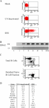
FIG. 1.
FIG. 1.
UV-inactivated EBV induces ISG expression in naive B cells. (A, B) Confirmation of EBV inactivation. 4E3 cells were infected with EBV-BXI from untreated or UV-inactivated stocks. (A) Cells were harvested at 24 h and analyzed by FACS. GFP expression levels were compared in mock-infected (top), UV-inactivated-EBV-infected (middle), and untreated-EBV-infected (lower) cells. (B) RNA was harvested from infected 4E3 cells, and RT-PCR was performed to detect transcript levels of EBNA2. (C) Naive B cells were separated from PBMC by negative selection. PBMC and the naive B-cell fractions were stained with PE-labeled anti-CD27 antibody (memory B-cell marker) and FITC-labeled anti-CD19 antibody (pan-B-cell marker) and analyzed by FACS. (D) Interferon-stimulated genes are induced in naive B cells by UV-inactivated and untreated EBV infection. RNA isolated from naive B cells 5 h after infection with UV- inactivated or untreated EBV was amplified by real-time PCR. Values are relative to those of uninfected naive B cells. Real-time assays were performed in triplicate, and results are representative of at least two experiments.
FIG. 2.
FIG. 2.
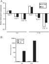
EBV induces TLR7. EBV infection induces TLR7 but represses TLR9 and -10 expression in naive B cells. Cells were infected with EBV (filled bars) or UV-inactivated EBV (open bars), and RNA was isolated at the indicated times after infection. (A) TLR expression. (B) EBV EBNA2 expression. RNA was quantified by real-time RT-PCR. The results are representative of two independent experiments performed in triplicate.
FIG. 3.
FIG. 3.
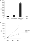
EBV-mediated stimulation of TLR7 enhances B-cell proliferation. (A) Naive B cells were incubated with UV-inactivated virus in the presence or absence of R837 (2 μg/ml) for 3 days. (B) Naive B cells were infected with BXI virus in the presence of a control oligonucleotide or IRS 661 (TLR7 inhibitory oligonucleotide) at 2.8 μM. Cells were treated at days 0, 3, and 5. Cell growth was quantitated in quadruplicate with Cell Titer Glo (Promega). Results are representative of two independent experiments. The standard deviation is shown. RLU, relative light units.
FIG. 4.
FIG. 4.
Expression of TLR7 and IRF-5 is associated with EBV primary infection, as well as type III latency. (A) TLR7 is upregulated in type III LCLs. Comparison of TLR7, TLR9, and TLR10 expression in the type III latently infected LCL cell line versus the EBV-negative 4E3 cell line. RNAs were quantified by real-time RT-PCR and gene-specific primers. The results are an average of two independent experiments performed in triplicate. (B, C) Induction of TLR7 after infection of EBV-negative 4E3 B cells. TLR7 (B) and EBV EBNA2 (C) RNAs were quantified by real-time RT-PCR. RNA was isolated at the indicated times after infection. The results are an average of two independent experiments performed in triplicate. (D) EBV induces IRF-5 transcripts. Shown are ethidium bromide-stained gels of electrophoretically separated RT-PCR products amplified from EBV-negative 4E3 B cells before (0 h) and at the indicated times after infection with Akata-BX1 virus. Primers were specific for IRF-5 (upper), EBV EBNA2 (middle), and TBP, which served as a loading control (lower). The results are representative of two independent experiments. (E) IRF-5 is expressed in type III latently infected cell lines. Western blot probed with anti-IRF-5 antibody comparing IRF-5 protein expression in type III latently infected cell lines (LCL, Mutu III, and EREB2-5), the EBV-positive EBNA2 deletion-containing cell line P3HR-1, a type I latently infected cell line (Mutu I), and EBV-negative B-cell lines (4E3 and Ramos). Equality of protein loading was determined by probing the membrane with anti-β-tubulin antibody.
FIG. 5.
FIG. 5.
EBV ameliorates negative growth effects following overstimulation of TLR7. Naive primary B cells were infected with EBV in medium without or with R837 (20 μg/ml) added initially at the time of infection (day 0) or 2 days postinfection (day 2) and subsequently supplemented on days 2 and 5. Cell proliferation was measured in quadruplicate with the Cell Titer Glo assay. The data shown are representative of two independent assays. The standard deviation is shown. RLU, relative light units.
FIG. 6.
FIG. 6.
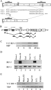
EBV infection induces a novel alternative splice variant of IRF-5, V12. (A) Exon structure of IRF-5 showing the predicted amino acid sequence of the variable exon 4 to 6 region of the V1, V3, and V12 cDNAs amplified by RT-PCR from EBV-infected cells. NLS, nuclear localization signal (3). NES, nuclear export signal (15, 44). DBD, DNA binding domain. TAD, transactivation domain. (B) Diagram of the structure of V12 cDNAs amplified by 5′ RLM RACE from Mutu III cells and cloned and sequenced. Shaded box, promoter region. (C to E) Ethidium bromide-stained, electrophoretically separated RT-PCR products amplified with primers specific for the V12 variant showing induction of V12 transcripts after EBV infection of Ramos cells (C) and expression of V12 transcripts in type III EBV-infected cell lines (Mutu III and LCL) but not in P3HR-1 or the type I latently infected B-cell lines (Mutu I, Rael) or gastric carcinoma cells (Snu719) (E). (D) Generic IRF-5 primers were used to amplify IRF-5 transcripts in the cell lines used in panel C. Amplification of cellular TBP or the ubiquitously expressed IRF-3 protein was used as a loading control. Results are representative of at least two experiments.
FIG. 7.
FIG. 7.
V12 IRF-5 is constitutively nuclear and negatively regulates IRF-5 transactivation. (A) V12 encodes a truncated IRF-5 protein. Western blot probed with anti-HA antibody comparing the electrophoretic mobility of polyacrylamide gel electrophoresis-separated, HA-tagged, in vitro-translated V3, V5, and V12 IRF-5 polypeptides. Lane C, control with no added template. NS, nonspecific bands. The values on the right are molecular sizes in kilodaltons. (B) Immunofluorescence assays comparing the intracellular localization of HA-tagged IRF-5 V3, V5, and V12 proteins and the phosphomimetic V3 protein (pV3) in transfected HeLa cells. Cells were stained with an anti-HA primary antibody and a rhodamine-labeled secondary antibody. Nuclei were stained with DAPI. (C) Reporter assay in which 293 cells were transfected with an IFNA1 promoter-luciferase reporter in the presence of V3 IRF-5, V12 IRF-5, pV3 IRF-5, or pV3 IRF-5 plus increasing amounts of V12 IRF-5 as indicated. The data shown are averages from two assays.
FIG. 8.
FIG. 8.
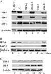
EBV induces expression of the IRF-5 negative regulator IRF-4. (A) Western blot assay comparing the expression of IRF-5 with that of IRF-4 and IRF-3 in different B-cell lines. Lane 1, 293 cells transfected with V3 IRF-5. Lanes 2, 4, and 6, type III latently infected cell lines. Lane 3, EBNA2 deletion-containing P3HR-1 cells. Lane 5, type I latently infected cell line. β-Tubulin served as a control for protein loading. (B) Western blot assay showing the dependence of IRF-4 expression on EBV latency gene expression. EREB2-5 cells were deprived of β-estradiol and harvested at the time points noted. Four days after withdrawal, β-estradiol was added back into the medium at 1 μM and cells were grown for a further 1 day (−4/+1). The membrane was probed with anti-IRF-4 and LMP1 antibodies. β-Tubulin was used as a loading control. (C) LMP-1 regulates IRF-4 expression. 1852, a tetracycline-on LMP-1, type III cell line, was grown in medium without tetracycline for 5 days, and then tetracycline was added back to the medium (time zero). A Western blot of the cell extract was probed with anti-IRF-4 and LMP1 antibodies. β-Tubulin was used as a loading control. The results are representative of two experiments.

References

    1. Akira, S., and K. Takeda. 2004. Toll-like receptor signalling. Nat. Rev. Immunol. 4:499-511. - PubMed
    1. Aman, P., and A. von Gabain. 1990. An Epstein-Barr virus immortalization associated gene segment interferes specifically with the IFN-induced anti-proliferative response in human B-lymphoid cell lines. EMBO J. 9:147-152. - PMC - PubMed
    1. Barnes, B. J., M. J. Kellum, A. E. Field, and P. M. Pitha. 2002. Multiple regulatory domains of IRF-5 control activation, cellular localization, and induction of chemokines that mediate recruitment of T lymphocytes. Mol. Cell. Biol. 22:5721-5740. - PMC - PubMed
    1. Barnes, B. J., M. J. Kellum, K. E. Pinder, J. A. Frisancho, and P. M. Pitha. 2003. Interferon regulatory factor 5, a novel mediator of cell cycle arrest and cell death. Cancer Res. 63:6424-6431. - "V体育ios版" PubMed
    1. Barnes, B. J., P. A. Moore, and P. M. Pitha. 2001. Virus-specific activation of a novel interferon regulatory factor, IRF-5, results in the induction of distinct interferon alpha genes. J. Biol. Chem. 276:23382-23390. - PubMed (VSports app下载)

Publication types

MeSH terms

Substances

LinkOut - more resources