Skip to main page content
U.S. flag

An official website of the United States government

Dot gov

The . gov means it’s official VSports app下载. Federal government websites often end in . gov or . mil. Before sharing sensitive information, make sure you’re on a federal government site. .

Https

The site is secure. The https:// ensures that you are connecting to the official website and that any information you provide is encrypted and transmitted securely V体育官网. .

Review
. 2007 Jul 15;463(2):134-48.
doi: 10.1016/j.abb.2007.04.013. Epub 2007 May 2.

Biochemical basis of regulation of human copper-transporting ATPases

Affiliations
Review

Biochemical basis of regulation of human copper-transporting ATPases

Svetlana Lutsenko (V体育平台登录) et al. Arch Biochem Biophys. .

Abstract

Copper is essential for cell metabolism as a cofactor of key metabolic enzymes. The biosynthetic incorporation of copper into secreted and plasma membrane-bound proteins requires activity of the copper-transporting ATPases (Cu-ATPases) ATP7A and ATP7B. The Cu-ATPases also export excess copper from the cell and thus critically contribute to the homeostatic control of copper VSports手机版. The trafficking of Cu-ATPases from the trans-Golgi network to endocytic vesicles in response to various signals allows for the balance between the biosynthetic and copper exporting functions of these transporters. Although significant progress has been made towards understanding the biochemical characteristics of human Cu-ATPase, the mechanisms that control their function and intracellular localization remain poorly understood. In this review, we summarize current information on structural features and functional properties of ATP7A and ATP7B. We also describe sequence motifs unique for each Cu-ATPase and speculate about their role in regulating ATP7A and ATP7B activity and trafficking. .


Figures

Figure 1
Figure 1. The dual role of copper-transporting ATPase ATP7B in hepatocyte
Copper enters the cell from the basolateral membrane via high-affinity copper transporter Ctr1 and is delivered to various cell targets with the help of copper chaperones. Atox1 transfers copper to ATP7B located in the TGN. ATP7B transports copper into the lumen of TGN, where copper is incorporated into ceruloplasmin (CP), which is subsequently excreted into the blood. When copper is elevated (red arrow), ATP7B traffics to vesicles. Vesicles filled with copper fuse with the apical (canalicular) membrane, copper is exported, and ATP7B is rapidly endocytosed. When copper is decreased ATP7B returns back to the TGN. It is possible that Atox1 regulates both copper delivery to ATP7B when copper is high and copper removal from ATP7B when copper is low.
Figure 2
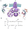
Figure 2. Trans-membrane organization and catalytic cycle of human Cu-ATPases
(A) Cartoon illustrating the major functional domains of Cu-ATPases. The N-terminal domain contains six copper-binding MBDs (MBD1–6). The transmembrane portion has eight TMS; the position of residues predicted to be involved in copper coordination (CPC, YN, and MxxS) is indicated. The A-domain may link changes in the N-terminal domain with those in the ATP-binding domain and in the transmembrane portion. The ATP-binding domain consists of the P-domain and the N-domain. The domains of ATP7B for which structure has been experimentally determined are indicated by dashed circles and corresponding structures are shown. Two Leu residues in the C-terminal tails required for endocytosis and/or return to TGN are indicated by “LL”. (B) The simplified catalytic cycle of human Cu-ATPases. Two major conformational states associated with high affinity for ATP and Cu (E1) and lower affinity for these ligands (E2) as well as phosphorylated intermediates (E1-Pi-Cu and E2-Pi-Cu) are shown
Figure 3
Figure 3. The sequence (A) and a predicted folding of transmembrane portion (B) for the transmembrane hairpin TMS1,2 of ATP7A and ATP7B
In (A), the residues conserved between ATP7A and ATP7B are high-lighted in grey; the putative TMS1 and TMS2 are underlined. The Met residues at the luminal end of TMS1 and TMS2 are in red; the putative metal-coordinating residues in the loop connecting TMS1 and TMS2 are in blue, the Cys residues in TMS1 and 2 are highlighted by yellow. In (B), the predicted structure of TMS1 and 2 without connecting loop is shown. The position of Cys residues is shown in yellow and the location of Met residues is in red.
Figure 4
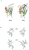
Figure 4. Metal binding pockets of N-terminal MBD2 and MBD3
(A) Comparison of the overall structure for the “typical” N-terminal domain, MBD2, and MBD3 of ATP7A according to (61,64) The corresponding secondary structure elements in two MBDs are indicated by the same color, the position of CxxC motif is indicated by red colors and letters. (B) The details of the metal binding pockets of N-terminal MBD2 and MBD3 in apo (left) and Cu-loaded (right) forms. Copper binding pocket of MBD2 of ATP7A (61) may be considered representative of the other MBDs of ATP7A and ATP7B. In MBD2, the loop containing GMxCxxC motif is flanked internally by the conserved Phe66. Copper binding to this region is associated with a small conformational shift (left). In the isolated MBD3 of ATP7A (58), Tyr69 performs the function of Phe66 and a larger rearrangement is necessary to coordinate copper. Copper coordinating cysteines are shown in green; copper is shown as a blue sphere. Letter R in the circles indicates side-chains non-coordinating amino-acid resides; they are shown to emphasize the rearrangement of the backbone upon copper binding
Figure 5
Figure 5. Schematic of N-Terminal domains of ATP7A (A) and ATP7B (B)
The length of inter-domain loops (in amino acid residues) is indicated. Loop is defined as the segment between folded 72-aa MBDs. (i) The loop between metal binding sites 1 and 2 of ATP7A is predicted to have a structure similar to the other metal binding sites, but lacks copper-coordinating residues. (ii) Metal binding sites 5 and 6 have been shown to be necessary for proper trafficking of ATP7A (87). (iii) The very N-Terminal region of ATP7A is also essential for proper trafficking and membrane targeting (69). (iv) MBS 2 has been shown to be the primary site of copper transfer from Atox1 (64). (v) MBS 4 has a higher affinity for Atox1 than does MBS2, and also has been shown capable of transferring copper to MBSs 5 and 6(57). However, in rat WNDP, this MBD lack CxxC motif and is not capable of copper binding. (vi) MBDs 5 and 6 are essential for copper transport, and have been shown to regulate affinity of the Intra-membrane copper binding site (40). (vii) Location of the G591D mutation, a disease causing mutation that disrupts Atox1 interactions with the N-Terminal domain.
Figure 6
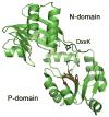
Figure 6. The ATP-binding domain of Cu-ATPases (based on the structure of CopA (35))
The invariant residues in proximity to the adenine moiety in the N-domain are indicated by the yellow color. The invariant Asp in the DKTG motif and the GDGxND sequence are marked in red. The position of residue equivalent to Asp1230 of ATP7A in the DxxK motif is in blue.

References

    1. Petris MJ, Strausak D, Mercer JF. Hum Mol Genet. 2000;9(19):2845–2851. - "V体育官网" PubMed
    1. Steveson TC, Ciccotosto GD, Ma XM, Mueller GP, Mains RE, Eipper BA. Endocrinology. 2003;144(1):188–200. - PubMed
    1. Tchaparian EH, Uriu-Adams JY, Keen CL, Mitchell AE, Rucker RB. Arch Biochem Biophys. 2000;379(1):71–77. - PubMed
    1. Qin Z, Itoh S, Jeney V, Ushio-Fukai M, Fukai T. Faseb J. 2006;20(2):334–336. - VSports手机版 - PubMed
    1. Terada K, Nakako T, Yang XL, Iida M, Aiba N, Minamiya Y, Nakai M, Sakaki T, Miura N, Sugiyama T. J Biol Chem. 1998;273(3):1815–1820. - PubMed

Publication types (V体育ios版)

MeSH terms