Skip to main page content
U.S. flag

An official website of the United States government

Dot gov

The . gov means it’s official. Federal government websites often end in . gov or . mil. Before sharing sensitive information, make sure you’re on a federal government site. VSports app下载.

Https

The site is secure. The https:// ensures that you are connecting to the official website and that any information you provide is encrypted and transmitted securely V体育官网. .

. 2007 Jul;27(13):4708-19.
doi: 10.1128/MCB.02432-06. Epub 2007 Apr 23.

Mammalian DET1 regulates Cul4A activity and forms stable complexes with E2 ubiquitin-conjugating enzymes

Affiliations

Mammalian DET1 regulates Cul4A activity and forms stable complexes with E2 ubiquitin-conjugating enzymes

Elah Pick et al. Mol Cell Biol. 2007 Jul.

Abstract

DET1 (de-etiolated 1) is an essential negative regulator of plant light responses, and it is a component of the Arabidopsis thaliana CDD complex containing DDB1 and COP10 ubiquitin E2 variant. Human DET1 has recently been isolated as one of the DDB1- and Cul4A-associated factors, along with an array of WD40-containing substrate receptors of the Cul4A-DDB1 ubiquitin ligase. However, DET1 differs from conventional substrate receptors of cullin E3 ligases in both biochemical behavior and activity. Here we report that mammalian DET1 forms stable DDD-E2 complexes, consisting of DDB1, DDA1 (DET1, DDB1 associated 1), and a member of the UBE2E group of canonical ubiquitin-conjugating enzymes. DDD-E2 complexes interact with multiple ubiquitin E3 ligases. We show that the E2 component cannot maintain the ubiquitin thioester linkage once bound to the DDD core, rendering mammalian DDD-E2 equivalent to the Arabidopsis CDD complex. While free UBE2E-3 is active and able to enhance UbcH5/Cul4A activity, the DDD core specifically inhibits Cul4A-dependent polyubiquitin chain assembly in vitro. Overexpression of DET1 inhibits UV-induced CDT1 degradation in cultured cells VSports手机版. These findings demonstrate that the conserved DET1 complex modulates Cul4A functions by a novel mechanism. .


Figures
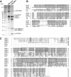
FIG. 1.
FIG. 1.
Isolation of human DET1 complex. (A) Silver staining of Flag-DET1 complex components isolated from HEK293 cells (mock) or Flag-DET1 stable cells. Asterisks indicate nonspecific bands. (B) Sequence alignment of DDA1 orthologs from various organisms. Homologous residues are shaded. (C) Sequence alignment of human UBE2E family E2s, including UBE2E-1 isoforms along with yeast UBC4 (yUBC4), Arabidopsis COP10, and UbcH5b and UbcH7. Identical residues are shaded. The conserved catalytic cysteine is indicated by an asterisk. Boxed sequences indicate the peptides identified by mass spectrometry and peptide sequencing.
FIG. 2.
FIG. 2.
Endogenous DET1 complex and specificity of DET1-associated E2s. (A) Endogenous DET1 and DDA1 were immunoprecipitated from HeLa cell extracts using their respective antisera or preimmune serum. The samples were immunoblotted using the indicated antibodies. (B) Whole-cell extract (input) and Flag-DET1 immunocomplex (IP-Flag) were analyzed by Western blotting using the indicated antibodies. (C) Recombinant GST-tagged UbcH5, UbE2E-1, UbE2E-2, UbE2E-3, and UbcH7 (50 ng per sample) were loaded on the SDS-PAGE gel. Anti-COP10 or anti-GST blots are shown. (D) Immunoprecipitations from Flag-DET1 cell extracts were performed using specific antibodies to UBE2E enzymes along with preimmune serum (pre-im) or other antibodies as indicated. The samples were analyzed by immunoblotting.
FIG. 3.
FIG. 3.
Characterization of DDA1. (A) His-DDA1 was coexpressed with untagged DDB1 (upper panel) or with T7-DET1 (lower panel) in E. coli. The elution from the Ni-NTA resin was examined by Coomassie brilliant blue staining (CBB) and anti-DDB1 or anti-T7 blot assays. (B) HeLa cells were transfected with Flag-DET1 or vector and were incubated with MG132 (10 μM) for 6 h in the indicated samples. Endogenous DDA1 was immunoprecipitated and analyzed by Western blotting. (C) DDA1 binds to the N-terminal domain of DDB1. Myc-tagged DDB1 and the truncation mutants were coexpressed with Flag-DDA1 in HEK293 cells. DDB1 proteins were pulled down by anti-myc resin, and association of Flag-DDA1 was detected by anti-Flag blotting. The data are summarized along with a diagram of the corresponding DDB1 derivatives in the bottom panel.
FIG. 4.
FIG. 4.
Characterization of the UBE2Es in DET1 complexes. (A and D) Superose-6 gel filtration analyses of total cell extracts from a Flag-DET1 stable line (A) or Flag-DET1 stable cell lines expressing myc-UBE2E-3N (D). Fractions were probed with the indicated antibodies. Fractions corresponding to CSN, DDD-E2, and free UBE2E-3 are labeled at the bottom. The myc-UBE2E-3N-containing DDD complex appeared slightly smaller than the endogenous DDD-UBE2E-3 complex. The Asterisk indicates the presumed partial breakdown product recognized by anti-UBE2E-3 but not by anti-myc antibody. (B) Flag-DET1 cell extracts were incubated with EDTA (5 mM) or increasing concentrations of ATP as indicated prior to Flag immunoprecipitation (IP). Decreased associations with UBE2E-3 and Cul4A were observed, while steady-state levels of UBE2E-3 and Cul4A did not change (not shown). (C) Myc-tagged UBE2E-3 full length (FL), UBE2E3-N(1-66) (N), or UBE2E3-C(60-207) (C) was transfected to Flag-DET1 cells. Total extract or Flag immunocomplex samples were blotted as indicated. (E) Proposed model describing the dynamic interactions of the UBE2Es and the DDD core complex.
FIG. 5.
FIG. 5.
(A) Coomassie blue gel of the rDDD complex consisting of HisT7-DET1, His-DDA1, and untagged DDB1. The complex was purified from E. coli by Ni-NTA resin. Identity of each component labeled on the right was verified by immunoblotting. (B) GST or GSTUBE2E-3 was preincubated under ubiquitin-loading conditions (E1+Ub + lanes) or not (- lanes) and then mixed with rDDD. Following GST pull-down, association of rDDD components was examined by immunoblotting. (C) Flag-DET1 cells were extracted in an ATP-containing buffer, followed by Flag immunoprecipitation (IP). Samples were denatured in a nonreducing (-DTT) or standard reducing (+DTT) sample buffer and probed with anti-UbcM2/UBE2E-3 (top) or anti-Flag (bottom). (D) Flag-DET1 cell extract containing 10 mM ATP was fractionated on a Superose-6 column. Fractions were mixed with nonreducing (top panel) or reducing (lower panels) sample buffer before immunoblotting. The ubiquitin-charged UBE2E-3 (UBE2E-3∼Ub) was found only in the “free” UBE2E-3 fractions. (E) In vitro ubiquitin thioester assay. Increasing amounts of recombinant GST-UBE2E-3 or Flag-DET1 immunocomplex were incubated with ubiquitin, E1, in an ATP-containing buffer in a 40-μl volume. The samples were probed with anti-UBE2E-3 antibody. The arrowhead indicates the ubiquitin thioester conjugate to UBE2E-3. The Flag-DET1 complex in lane 2 contained about 70 ng of UBE2E-3 as estimated by silver staining.
FIG. 6.
FIG. 6.
DDD-E2 complex association with E3 ligases and its effect on Cul4A E3 activity. (A) Whole-cell extract (total) or Flag-DET1 immunocomplex (IP-Flag) was analyzed by Western blotting using the indicated antibodies. (B) His-ARA54 was transiently expressed in Flag-DET1 cells. Proteins in the Ni-NTA pull-down were blotted for anti-His and anti-Flag. (C) All reaction mixtures contained ubiquitin, biotinylated ubiquitin, and E1. UbcH5c (50 ng) or Flag-DET1 complexes containing approximately (50 ng) of E2s were used as E2 sources. (D) UBE2E-3 is an active E2 for Cul4A. GST, GST-UBE2E-3, or its catalytic site C/S mutant (2.5 ng/μl) was used as E2. Ubiquitin chains were detected by antiubiquitin blotting. Equal amounts of Cul4A and GST fusion proteins were confirmed by immunoblotting (lower two panels). (E) A similar experiment was set up as for the experiment in panel D, except that the E2s were UbcH5 (2.5 ng/μl) in combination with GST (5 ng/μl; lanes 1 and 2), UBE2E-3 (5 ng/μl; lanes 5 and 6), or UBE2E-3(C/S) mutant (5 ng/μl; lanes 3 and 4). UBE2E-3(C/S) enhanced polyubiquitination when combined with UbcH5 and Cul4A, while addition of rDDD complex (50 ng/μl) strongly inhibited polyubiquitination detected by antiubiquitin blotting (upper panel). Amounts of UBE2E-3, Cul4A, and DET1 were confirmed by anti-GST and anti-T7 blotting.
FIG. 7.
FIG. 7.
In vitro E3 activity assay using reconstituted CRL E3. (A) Recombinant Cul4A-DDB1-Rbx1 complex (2 μg) or (B) recombinant Cul1(324-776)-Roc1 complex (0.3 μg) was incubated in a 15-μl reaction mixture containing ubiquitin, ATP, E1, and UbcH5b (0.2 μg). As indicated, bovine serum albumin (BSA; 1.25 and 2.5 μg) or the following recombinant proteins that had equal amounts of DDA1 were added to the reaction mixture: His-DDA1, DDB1 (0.6 and 1.2 μg); His-DDA1, T7-DET1 (0.75 and 1.5 μg); His-DDA1, HisT7-DET1, and DDB1 (rDDD, 0.75 and 1.5 μg). Ubiquitin chain was detected by antiubiquitin blotting. Total protein amounts in the reaction mixtures were examined by Ponceau S staining of the membrane. Identity of labeled protein bands was confirmed by immunoblotting. (C and D) Experiments similar to those in panels A and B, except that the recombinant complexes were normalized for equal amounts of DET1 in the reaction: His-DDA1, DDB1 (3 and 6 μg); His-DDA1, T7-DET1 (5.5 and 11 μg); His-DDA1, HisT7-DET1, DDB1 (rDDD, 0.75 and 1.5 μg). Amounts of DET1 were confirmed by anti-T7 blotting. Ubiquitin chain was detected by antiubiquitin blotting.
FIG. 8.
FIG. 8.
DET1 inhibits UV-stimulated degradation of CDT1. (A) HeLa cells in 12-well plates were transfected with siRNA oligos corresponding to Lamin A (control), DET1, DDA1, and a combination of UBE2E-1, -2, and -3. After 48 h, cells were irradiated with UV-C light (40 J/m2). Samples were collected 15 min after the UV treatment by direct lysis. (B) HeLa cells in 12-well plates were transfected with 4 μg of the plasmids expressing different DDD-E2 subunits. After 24 h, cells were exposed to UV-C light (40 J/m2) and collected by direct lysis in SDS sample buffer at the time points indicated. Levels of CDT1 and overexpressed proteins were detected by immunoblotting. An asterisk denotes a nonspecific band cross-reacting with the anti-CDT1 antibody.

V体育2025版 - References

    1. Angers, S., T. Li, X. Yi, M. J. Maccoss, R. T. Moon, and N. Zheng. 2006. Molecular architecture and assembly of the DDB1-CUL4A ubiquitin ligase machinery. Nature 443:590-593. - PubMed
    1. Benvenuto, G., F. Formiggini, P. Laflamme, M. Malakhov, and C. Bowler. 2002. The photomorphogenesis regulator DET1 binds the amino-terminal tail of histone H2B in a nucleosome context. Curr. Biol. 12:1529-1534. - PubMed (V体育官网)
    1. Bernhardt, A., E. Lechner, P. Hano, V. Schade, M. Dieterle, M. Anders, M. J. Dubin, G. Benvenuto, C. Bowler, P. Genschik, and H. Hellmann. 2006. CUL4 associates with DDB1 and DET1 and its downregulation affects diverse aspects of development in Arabidopsis thaliana. Plant J. 47:591-603. - PubMed
    1. Bondar, T., E. V. Mirkin, D. S. Ucker, W. E. Walden, S. M. Mirkin, and P. Raychaudhuri. 2003. Schizosaccharomyces pombe Ddb1 is functionally linked to the replication checkpoint pathway. J. Biol. Chem. 278:37006-37014. - PubMed (VSports手机版)
    1. Chen, H., Y. Shen, X. Tang, L. Yu, J. Wang, L. Guo, Y. Zhang, H. Zhang, S. Feng, E. Strickland, N. Zheng, and X. W. Deng. 2006. Arabidopsis CULLIN4 forms an E3 ubiquitin ligase with RBX1 and the CDD complex in mediating light control of development. Plant Cell 18:1991-2004. - PMC - PubMed

Publication types

V体育安卓版 - MeSH terms