Skip to main page content
U.S. flag

An official website of the United States government

Dot gov

The . gov means it’s official. Federal government websites often end in . gov or . mil. Before sharing sensitive information, make sure you’re on a federal government site VSports app下载. .

Https

The site is secure. The https:// ensures that you are connecting to the official website and that any information you provide is encrypted and transmitted securely V体育官网. .

. 2007 Apr 15;21(8):942-55.
doi: 10.1101/gad.1528507.

Single-stranded DNA-binding proteins regulate the abundance of LIM domain and LIM domain-binding proteins

Affiliations

V体育2025版 - Single-stranded DNA-binding proteins regulate the abundance of LIM domain and LIM domain-binding proteins

Zhixiong Xu et al. Genes Dev. .

Abstract

The LIM domain-binding protein Ldb1 is an essential cofactor of LIM-homeodomain (LIM-HD) and LIM-only (LMO) proteins in development. The stoichiometry of Ldb1, LIM-HD, and LMO proteins is tightly controlled in the cell and is likely a critical determinant of their biological actions. Single-stranded DNA-binding proteins (SSBPs) were recently shown to interact with Ldb1 and are also important in developmental programs. We establish here that two mammalian SSBPs, SSBP2 and SSBP3, contribute to an erythroid DNA-binding complex that contains the transcription factors Tal1 and GATA-1, the LIM domain protein Lmo2, and Ldb1 and binds a bipartite E-box-GATA DNA sequence motif. In addition, SSBP2 was found to augment transcription of the Protein 4. 2 (P4. 2) gene, a direct target of the E-box-GATA-binding complex, in an Ldb1-dependent manner and to increase endogenous Ldb1 and Lmo2 protein levels, E-box-GATA DNA-binding activity, and P4. 2 and beta-globin expression in erythroid progenitors. Finally, SSBP2 was demonstrated to inhibit Ldb1 and Lmo2 interaction with the E3 ubiquitin ligase RLIM, prevent RLIM-mediated Ldb1 ubiquitination, and protect Ldb1 and Lmo2 from proteasomal degradation. These results define a novel biochemical function for SSBPs in regulating the abundance of LIM domain and LIM domain-binding proteins VSports手机版. .


Figures
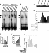
Figure 1.
Figure 1.
Ssbp2 and Ssbp3 are integral components of an erythroid E-box-GATA DNA-binding complex. (A) EMSA of nuclear extracts from MEL and FVA cells for E-box-GATA DNA-binding activity. Rabbit IgG or antibodies to GATA-1, Ldb1, or SSBP2 were used as indicated for supershift analysis. Complexes supershifted by antibodies to GATA-1 and Ldb1 are marked with solid arrowheads and complexes containing only GATA-1 are marked with filled circles. Complexes supershifted by SSBP2 antibody are marked to the right of the relevant lane with asterisks. (B) EMSA of nuclear extracts from MEL cells for E-box-GATA DNA-binding activity. Normal rabbit IgG or antibodies to SSBP3 and SSBP2 were used for supershift analysis and DNA-binding complexes marked as above. (C) ChIP analysis of SSBP2 and Ldb1 occupancy at the P4.2 promoter in differentiating MEL cells. (D) Transient transfection analysis with a plasmid (1.4 μg) containing 1.7 kb of the P4.2 promoter linked to a luciferase reporter gene in COS-7L cells transfected with (filled column) or without (unfilled column) indicated combinations of expression plasmids pcDNA3.1-Tal1 (100 ng), pcDNA3.1-E47 (3 ng), pcDNA3.1-GATA-1 (25 ng), pEFIRES-Ldb1 (40 ng), pEFIRES-Ldb1(Δ214–223) (40 ng), pEFIRES-Lmo2 (40 ng), pEFIRES-SSBP2 (200 ng), pEFIRES-SSBP2(Δ95–261) (200 ng), and pEFIRES-SSBP2(Δ51–94) (200 ng). The mean ± SEM luciferase activity relative to that with reporter alone from three independent experiments is plotted. Reporter analysis in DMSO-treated MEL cells transfected with (filled column) or without (unfilled column) indicated amounts of pSilencer-Ssbp2-B (E), pEFIRES-SSBP2 (F), and indicated combinations of pEFIRES-SSBP2 (500 ng), pEFIRES-SSBP2(Δ95–261) (500 ng), pEFIRES-SSBP2(Δ51–94) (500 ng), pEFIRES-Ldb1 (500 ng), and pEFIRES-Ldb1(Δ214–223) (500 ng) (G).
Figure 2.
Figure 2.
SSBP2 positively regulates P4.2 and β-globin gene expression in MEL cells. (A) Western blot analysis of Flag-tagged SSBP2 and SSBP2(Δ95–261) mutant in MEL whole cellular extracts with antibody to the Flag epitope (top) and nuclear extracts with antibody to amino acids 269–284 of SSBP2 (bottom). A cross-reacting protein recognized by Flag antibody is marked with an asterisk and served as loading control. (B) Quantitative RT–PCR analysis of P4.2, β-globin, and RLIM mRNAs in indicated MEL cell transductants. The mean ± SEM RNA level relative to vector control is plotted.
Figure 3.
Figure 3.
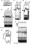
Overexpression of SSBP2 increases E-box-GATA DNA-binding activity and P4.2 promoter occupancy in MEL cells. (A) Western blot analysis of Ssbp2 (top) and Ssbp3 (bottom) protein expression in whole cellular extracts of differentiating MEL cells at times after DMSO addition. Actin was used as loading control. (B) EMSA of E-box-GATA DNA-binding activity in nuclear extracts from SSBP2-transduced MEL cells. The upper ternary complex is marked with an open arrowhead, and the lower complex, whose abundance was stimulated by SSBP2 overexpression, is marked with a solid arrowhead. The levels of DNA-binding activity for the lower complex were normalized to a nonspecific protein–DNA complex (asterisk). The fold change relative to vector control is shown below the autoradiographs. Antibody supershift analysis of nuclear extracts from oligoclonal population O4 transduced with full-length SSBP2 (C) or a polyclonal population transduced with SSBP2(Δ95–261) (D) using antibodies to Flag epitope, Ldb1, and GATA-1. DNA-binding complexes are marked as above. (E) Quantitative ChIP analysis of factor occupancy at the promoter or 3′ UTR of the P4.2 gene for vector-transduced (unfilled column) and SSBP2-transduced (filled column) MEL cells. The mean ± SEM occupancy as a percentage of input for SSBP2, Ldb1, and GATA-1 is plotted. (F) EMSA of E-box-GATA DNA-binding activity using nuclear extracts (5 μg) from COS-7L cells transfected with indicated combinations of pEFIRES-SSBP2, pEFIRES-Ldb1, and pEFIRES-Lmo2, assayed as mixed with nuclear extract (5 μg) from undifferentiated MEL cells. The levels of DNA-binding activity of the lower complex were normalized as above. The fold change relative to vector control is shown below the autoradiographs.
Figure 4.
Figure 4.
SSBP2 regulates the abundance of Ldb1 and Lmo2 proteins. (A) Western blot analysis of Tal1, GATA-1, Hdac2, Ldb1, and Lmo2 abundance in nuclear extracts from transduced MEL cells. The levels of Ldb1 and Lmo2 in SSBP2- or SSBP2(Δ95–261)-transduced cells relative to vector control are shown below the autoradiographs. (B) Western blot analysis of endogenous Ldb1 and Hdac2 expression and transduced Flag-tagged SSBP2 protein expression in COS-7L and CHO cells transiently transfected with increasing amounts of pEFIRES-SSBP2 (175 ng, 525 ng, and 1.6 μg for COS-7L cells; 125 ng and 1.0 μg for CHO cells). (C) Quantitative RT–PCR analysis of Ldb1 expression in indicated MEL cell transductants. The mean ± SEM RNA level relative to that in vector control is plotted. (D) Protein turnover analysis in SSBP2-transduced MEL cells treated with 100 μM CHX for the indicated times. (Top) Ldb1 expression was detected by Western blot analysis and quantified by densitometry of X-ray films. (Bottom) The amount of protein remaining is expressed as a function of time from addition of CHX.
Figure 5.
Figure 5.
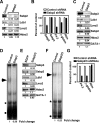
Depletion of Ssbp2 and Ssbp3 reduces Ldb1 and Lmo2 abundance and E-box-GATA DNA-binding activity. (A) Western blot analysis of Ssbp2, Ldb1, Lmo2, and Hdac2 expression in whole cellular extracts from Ssbp2 shRNA-transduced MEL cells treated with 1.5% DMSO for 2 d. The levels of proteins in cells expressing Ssbp2 shRNA relative to those expressing a control shRNA are shown below the autoradiographs. (B) Quantitative RT–PCR analysis of Ssbp2, Ssbp3, P4.2, β-globin, and Rlim mRNAs in differentiating MEL cells transiently transfected with expression plasmid for Ssbp2 shRNA or control shRNA. (C) Western blot analysis of Ssbp3, Ldb1, Lmo2, Ssbp2, Hdac2, and GATA-1 expression in nuclear extracts from short-term puromycin-selected MEL cells transfected with expression plasmid for Ssbp3 shRNA or control shRNA. (D) EMSA of nuclear extracts prepared as above. The E-box-GATA DNA-binding complex is marked with a solid arrowhead. Its abundance was normalized to that of a nonspecific protein–DNA complex (asterisk). The fold change in E-box-GATA DNA-binding activity in cells expressing Ssbp3 shRNA relative to those expressing control shRNA is shown below the autoradiographs. (E) Western blot analysis of Ssbp3, Ssbp2, Ldb1, Lmo2, Hdac2, and GATA-1 abundance in nuclear extracts from short-term puromycin-selected MEL cells expressing shRNAs to both Ssbp2 and Ssbp3 or control (EGFP) shRNA. (F) EMSA of nuclear extracts prepared as above. Protein–DNA complexes are marked as in D. (G) Quantitative RT–PCR analysis of P4.2, β-globin, and Rlim mRNAs in differentiating MEL cells transfected with shRNA expression plasmids for Ssbp2 and Ssbp3 or a control sequence (EGFP).
Figure 6.
Figure 6.
SSBP2 prevents RLIM-mediated degradation of Ldb1 and Lmo2 proteins. (A) Western blot analysis of CHO cells transfected with indicated combinations of pTracer-EF/V5-Ldb1 (400 ng), pTracer-EF/V5-Ldb1(Δ214–223) (400 ng), pCMV6-XL4-RLIM (1.2 μg), and pEFIRES-SSBP2 (100, 200, or 400 ng). The total mass of DNA was adjusted to 2 μg with plasmid pCMV4 as needed. Whole cellular extracts were prepared 48 h after transfection and assayed for protein expression using a monoclonal antibody to the V5 epitope and polyclonal antibody to the Flag epitope. A cross-reacting protein recognized by the Flag antibody is marked with an asterisk and served as loading control. (B) Western blot analysis of CHO cells transfected with indicated combinations of pTracer-EF/V5-Ldb1 (300 ng), pTracer-EF/V5-Ldb1(Δ214–223) (300 ng), pTracer-EF/V5-Lmo2 (500 ng), pCMV6-XL4-RLIM (800 ng), and pEFIRES-SSBP2 (400 ng). V5- and Flag-tagged proteins were detected as above. (C) Western blot analysis of CHO cells transfected with indicated combinations of pTracer-EF/V5-Ldb1 (300 ng), pTracer-EF/V5-Lmo2 (500 ng), pCMV6-XL4-RLIM (800 ng), pEFIRES-SSBP2 (100, 200, or 400 ng), pEFIRES-SSBP2(Δ95–261) (400 ng), SSBP2(Δ51–94) (400 ng), and SSBP2(1–94) (400 ng). (D) Western blot analysis of CHO cells transfected as above using indicated combinations of pTracer-EF/V5-Ldb1b (300 ng), pTracer-EF/V5-Lmo2 (500 ng), pTracer-EF/V5-RLIM (800 ng), and pEFIRES-SSBP2 (400 ng). (E) Western blot analysis of CHO cells transfected as in C with pTracer-EF/V5-LMO4 instead of pTracer-EF/V5-Lmo2.
Figure 7.
Figure 7.
SSBP2 inhibits RLIM-mediated Ldb1 ubiquitination and RLIM interaction with Ldb1. (A) Western blot analysis of CHO cells transfected with indicated combinations of pEFIRES-Ldb1 (400 ng), pCMV6-XL4-RLIM (1.0 μg), pEFIRES-SSBP2 (300 ng), pEFIRES-SSBP2(1–94) (300 ng), and CMV-HA-ubiquitin (300 ng). Whole cellular extracts prepared from MG132-treated transfectants were subjected to coimmunoprecipitation analysis as indicated. (B) Lack of deubiquitinating activity of SSBP2. Equivalent amounts of ubiquitinated Ldb1 immobilized on beads were incubated with or without purified Flag-SSBP2 protein. Ubiquitinated Ldb1 and Flag-SSBP2 retained on beads were then quantified by Western blot analysis. (C) Western blot analysis of CHO cells transfected as above with indicated combinations of pEFIRES-Ldb1 (500 ng), pTracer-EF/V5-Lmo2 (500 ng), pTracer-EF/V5-RLIM (500 ng), pEFIRES-SSBP2 (500 ng), and pEFIRES-SSBP2(1–94) (500 ng). Whole cellular extracts from MG132-treated transfectants were subjected to coimmunoprecipitation analysis as indicated. Western blot analysis of SSBP2, Ldb1, and Lmo2 in purified SSBP2 protein complexes was carried out following denaturing (D) and nondenaturing (E) polyacrylamide gel electrophoresis. CHO cells were transfected with expression plasmids for Ldb1, Lmo2, and Flag-tagged SSBP2, and Western blot analysis of SSBP2-associated proteins was carried out on purified complexes following elution with Flag peptide.
Figure 8.
Figure 8.
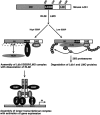
Model of SSBP regulation of Ldb1 and LMO protein stability and transcriptional complex assembly. Top shows schematic of mouse Ldb1 with dimerization domain (DD), Ldb1/Chip conserved domain (LCCD), and LIM interaction domain (LID) marked. The model predicts that when at sufficient levels, SSBPs interact with Ldb1 to promote formation of an SSBP/Ldb1/LMO trimolecular complex, with Ldb1 contacting SSBP and Lmo2 simultaneously through its separate LCCD and LID, respectively. This prevents their interaction with the ubiquitin ligase RLIM, RLIM-initiated ubiquitination, and proteasomal degradation. The SSBP/Ldb1/LMO complex could also help nucleate assembly of larger transcriptional complexes, including the E-box-GATA DNA-binding complex shown. When SSBPs are present at insufficient levels to interact with Ldb1, RLIM promotes the ubiquitination of Ldb1 and LIM-only proteins, targeting them for destruction in the 26S proteasome.

References

    1. Agulnick A.D., Taira M., Breen J.J., Tanaka T., Dawid I.B., Westphal H., Taira M., Breen J.J., Tanaka T., Dawid I.B., Westphal H., Breen J.J., Tanaka T., Dawid I.B., Westphal H., Tanaka T., Dawid I.B., Westphal H., Dawid I.B., Westphal H., Westphal H. Interactions of the LIM-domain-binding factor Ldb1 with LIM homeodomain proteins. Nature. 1996;384:270–272. - VSports最新版本 - PubMed
    1. Amerik A.Y., Hochstrasser M., Hochstrasser M. Mechanism and function of deubiquitinating enzymes. Biochim. Biophys. Acta. 2004;1695:189–207. - PubMed
    1. Aoyama M., Ozaki T., Inuzuka H., Tomotsune D., Hirato J., Okamoto Y., Tokita H., Ohira M., Nakagawara A., Ozaki T., Inuzuka H., Tomotsune D., Hirato J., Okamoto Y., Tokita H., Ohira M., Nakagawara A., Inuzuka H., Tomotsune D., Hirato J., Okamoto Y., Tokita H., Ohira M., Nakagawara A., Tomotsune D., Hirato J., Okamoto Y., Tokita H., Ohira M., Nakagawara A., Hirato J., Okamoto Y., Tokita H., Ohira M., Nakagawara A., Okamoto Y., Tokita H., Ohira M., Nakagawara A., Tokita H., Ohira M., Nakagawara A., Ohira M., Nakagawara A., Nakagawara A. LMO3 interacts with neuronal transcription factor, HEN2, and acts as an oncogene in neuroblastoma. Cancer Res. 2005;65:4587–4597. - PubMed (VSports手机版)
    1. Bach I., Carriere C., Ostendorff H.P., Andersen B., Rosenfeld M.G., Carriere C., Ostendorff H.P., Andersen B., Rosenfeld M.G., Ostendorff H.P., Andersen B., Rosenfeld M.G., Andersen B., Rosenfeld M.G., Rosenfeld M.G. A family of LIM domain-associated cofactors confer transcriptional synergism between LIM and Otx homeodomain proteins. Genes & Dev. 1997;11:1370–1380. - PubMed
    1. Bayarsaihan D., Lukens L.N., Lukens L.N. Single-strand-DNA-binding factors specifically recognize the pyrimidine element in the chick α2(I) collagen gene promoter. Biochem. J. 1996;314:293–296. - PMC - PubMed

Publication types

MeSH terms

"VSports手机版" LinkOut - more resources