Skip to main page content (V体育2025版)
U.S. flag

An official website of the United States government

Dot gov

The . gov means it’s official. Federal government websites often end in . gov or . mil VSports app下载. Before sharing sensitive information, make sure you’re on a federal government site. .

Https

The site is secure. The https:// ensures that you are connecting to the official website and that any information you provide is encrypted and transmitted securely V体育官网. .

. 2007 May;117(5):1305-13.
doi: 10.1172/JCI30740. Epub 2007 Apr 5.

Inhibition of TGF-beta with neutralizing antibodies prevents radiation-induced acceleration of metastatic cancer progression

Affiliations

Inhibition of TGF-beta with neutralizing antibodies prevents radiation-induced acceleration of metastatic cancer progression

Swati Biswas et al. J Clin Invest. 2007 May.

Erratum in

Abstract

We investigated whether TGF-beta induced by anticancer therapies accelerates tumor progression. Using the MMTV/PyVmT transgenic model of metastatic breast cancer, we show that administration of ionizing radiation or doxorubicin caused increased circulating levels of TGF-beta1 as well as increased circulating tumor cells and lung metastases. These effects were abrogated by administration of a neutralizing pan-TGF-beta antibody VSports手机版. Circulating polyomavirus middle T antigen-expressing tumor cells did not grow ex vivo in the presence of the TGF-beta antibody, suggesting autocrine TGF-beta is a survival signal in these cells. Radiation failed to enhance lung metastases in mice bearing tumors that lack the type II TGF-beta receptor, suggesting that the increase in metastases was due, at least in part, to a direct effect of TGF-beta on the cancer cells. These data implicate TGF-beta induced by anticancer therapy as a pro-metastatic signal in tumor cells and provide a rationale for the simultaneous use of these therapies in combination with TGF-beta inhibitors. .


"VSports最新版本" Figures

Figure 1
Figure 1. Radiation and chemotherapy increase circulating TGF-β1.
(A) FVB mice were subjected to 10 Gy delivered to the thorax (left) or pelvis (right). Blood was collected 24 hours later, and plasma TGF-β1 level was measured as described in Methods. (B) Eight-week-old tumor-bearing MMTV/PyVmT mice or nontransgenic FVB mice bearing PyVmT tumors of 200 mm3 or greater in mammary fat pad no. 4 were left untreated or administered 10 Gy to the thorax. Plasma TGF-β1 levels were measured 24 hours later. (C) Transgenic mice were treated 3 times with vehicle or doxorubicin (5 mg/kg i.p.) at 21-day intervals starting at week 8. TGF-β1 was measured in plasma collected at week 15. Data in AC represent 3 independent experiments using 3 subjects per group. (D) FVB mice were administered 10 Gy to the thorax. Five weeks later, lungs from irradiated mice and controls were harvested and lysates (250 μg/ml) added in triplicate wells to mink lung epithelial cells that stably express a plasminogen activator inhibitor–1/luciferase reporter (PAI-1/luciferase reporter). After 24 hours, luciferase expression was measured as described in Methods. *P < 0.05, **P < 0.01, ***P < 0.001 versus control.
Figure 2
Figure 2. Radiation and chemotherapy increase circulating tumor cells and lung metastases.
(A) Female MMTV/PyVmT female mice were subjected to thoracic irradiation at 8 weeks of age. At 13 weeks, blood was collected via heart puncture at the termination of the experiment, and its cellular fraction evaluated for its ability to form colonies ex vivo as described in Methods. CTCs, circulating tumor cells. Representative images of colonies arising from single circulating tumor cells harvested from mouse blood are shown below. Transgene-positive colonies were assessed under a fluorescence microscope using a PyVmT antibody and a fluorescent secondary antibody. (B) In same mice as in A, surface lung metastases were counted at 13 weeks of age. Data in A and B are representative of 3 independent experiments with 4 mice per group. Representative H&E-stained sections of lung tissues from control and irradiated transgenic mice obtained 5 weeks after thoracic radiation are shown below. Black arrows indicate lung metastases. (C) Eight-week-old tumor-bearing MMTV/PyVmT transgenic mice were treated 3 times with vehicle or doxorubicin (5 mg/kg i.p.) at 21-day intervals. The experiment was terminated on week 15, and surface lung metastases were counted. Representative H&E-stained lung sections containing metastatic foci are shown at right. Original magnification, ×100. *P < 0.05, **P < 0.01 versus control.
Figure 3
Figure 3. Thoracic radiation increases lung metastases from tumor transplants.
(A) MMTV/PyVmT cells stably expressing luciferase were injected in the mammary fat pads of syngeneic FVB mice. Bioluminescence imaging was used to monitor tumor growth and lung metastases twice a week thereafter. A representative mouse imaged 2 weeks after cell inoculation is shown. (B and C) Mice with PyVmT/Luc tumors measuring at least 200 mm3 were treated or not with thoracic irradiation (10 Gy). (B) Surface lung metastases were quantitated 2 weeks later. Data are mean ± SD of 5 mice per group in 1 of 2 experiments. **P < 0.01 versus control. (C) H&E slides of lung and primary tumor sections. Original magnification, ×100.
Figure 4
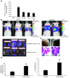
Figure 4. Prior radiation is permissive for metastatic lung colonization in tumor-free mice.
(A) MMTV/PyVmT cells in 100-mm dishes in serum-free medium were treated with 1.25–7.5 Gy. Cell-conditioned medium was collected 72 hours later, and TGF-β1 levels were determined by ELISA as described in Methods. (B) MMTV/PyVmT cells stably expressing luciferase were injected via tail vein in virgin female FVB mice. Where indicated, mice received 10 Gy to the thorax 1 hour prior to injection of cells. Cancer cells in lungs were visualized by mouse bioluminescence 2 weeks after inoculation (top). In some cases, lungs were surgically removed after administration of d-luciferin and imaged ex vivo (bottom). Controls are shown on the left and irradiated mice are shown on the right. (C) Representative lung whole mounts (top) and H&E sections of lungs (bottom; original magnification, ×100) from control and irradiated mice. (D) Quantification of surface lung metastasis (left) and lung weight (right) in control and irradiated mice. Data are mean ± SD of 5 mice per group in 2 independent experiments. **P < 0.01, ***P < 0.001 versus control.
Figure 5
Figure 5. TGF-β–neutralizing antibody 2G7 blocks radiation-induced increase in lung metastases.
(A and B) Eight-week-old tumor-bearing MMTV/PyVmT mice were administered 10 Gy to the thorax. Where indicated, mice were treated with 15 mg/kg of 2G7 twice a week until week 13, at which time surface lung metastases were counted (A). Data are mean ± SD of 5 mice per group. (B) Representative H&E stains of lung sections. The experiment was repeated once with similar results. (C) Blood was collected at the completion of the experiment via heart puncture and its cellular fraction evaluated for its ability to form colonies ex vivo as described in Methods. (D) At 13 weeks, blood was collected from tumor-bearing transgenic mice that were exposed to thorax irradiation. The cellular fraction was plated ex vivo as in C in the presence of 20 μg/ml 2G7 or PBS. Colonies measuring 50 μm or greater were counted manually 10–12 days later. Data are mean ± SD of 5 mice per group. *P < 0.05, **P < 0.01 versus control.
Figure 6
Figure 6. Absence of TGFβRII in tumor cells abrogates radiation-induced increase in lung metastases.
(AC) PyVmT/TGFBR2flox/flox and PyVmT/TGFBR2KO cells stably expressing luciferase were injected via the tail vein in virgin 8-week-old female FVB mice that had received or not 10 Gy to the thorax 1 hour prior to tumor cell injection. Two weeks later, surface lung metastases were evaluated by bioluminescence (A), by histology (B), and by manually counting surface lung metastases (C) as described in Methods. Data are mean ± SD of 4 mice per group. (D) PCR from genomic DNA extracted from both cell lines, showing the presence of a recombined band only in PyVmT/TGFBR2KO cells. **P < 0.01 versus control.

References

    1. Massague J., Seoane J., Wotton D. Smad transcription factors. Genes Dev. 2005;19:2783–2810. - PubMed
    1. Siegel P.M., Massague J. Cytostatic and apoptotic actions of TGF-beta in homeostasis and cancer. Nat. Rev. Cancer. 2003;3:807–820. - PubMed
    1. Bierie B., Moses H.L. Tumour microenvironment: TGFbeta: the molecular Jekyll and Hyde of cancer. Nat. Rev. Cancer. 2006;6:506–520. - PubMed
    1. Dumont N., Arteaga C.L. Targeting the TGF beta signaling network in human neoplasia. Cancer Cell. 2003;3:531–536. - "VSports最新版本" PubMed
    1. Wakefield L.M., Roberts A.B. TGF-beta signaling: positive and negative effects on tumorigenesis. Curr. Opin. Genet. Dev. 2002;12:22–29. - "V体育官网" PubMed

Publication types (V体育2025版)

V体育2025版 - MeSH terms

Substances