"V体育平台登录" Skip to main page content
U.S. flag

An official website of the United States government

Dot gov

The . gov means it’s official. Federal government websites often end in . gov or . mil VSports app下载. Before sharing sensitive information, make sure you’re on a federal government site. .

Https

The site is secure V体育官网. The https:// ensures that you are connecting to the official website and that any information you provide is encrypted and transmitted securely. .

. 2007 Apr 15;178(8):5028-34.
doi: 10.4049/jimmunol.178.8.5028.

Beta-catenin regulates positive selection of thymocytes but not lineage commitment

Affiliations

Beta-catenin regulates positive selection of thymocytes but not lineage commitment

Qing Yu et al. J Immunol. .

Abstract

Positive selection and lineage commitment to the cytolytic or helper lineage of T cells result in coordinated expression of MHC class I-restricted TCR and CD8 coreceptor or MHC class II-restricted TCR and CD4 molecule. Positive selection signals also regulate the survival and generation of requisite numbers of cytolytic or Th cells. beta-Catenin is the major transcriptional cofactor of T cell factor and plays a role in thymocyte development VSports手机版. In this study, using mice expressing stabilized beta-catenin and mice with T cell-specific deletion of beta-catenin, we show that beta-catenin regulates positive selection, but not lineage commitment of thymocytes. Furthermore, beta-catenin expression accelerates the timing of mature CD8 thymocyte generation such that CD4 and CD8 single-positive thymocytes mature with the same kinetics during development. .


Figures

FIGURE 1
FIGURE 1
Intrathymic signals regulate stabilization of β-catenin in a developmentally significant manner. A, Expression of endogenous (wild-type) and Tg β-catenin protein. Whole cell lysates from sorted thymocyte subpopulations from control C57BL/6 mice or CAT-Tg mice were subjected to Western blot analysis with anti-β-catenin or anti-protein kinase Cμ Ab. Data are representative of three independent experiments. B, Real-time RT-PCR for mRNA for endogenous β-catenin. Sorted thymocyte subpopulations from control C57BL/6 mice or CAT-Tg mice were subjected to real-time RT-PCR for expression of endogenous β-catenin gene. Relative expression of β-catenin to β-actin is normalized with that in control DN thymocytes set as 1 (n = 4).
FIGURE 2
FIGURE 2
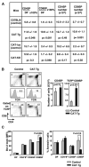
β-Catenin expression increases the number of CD8SP thymocytes. A, Percentage and number of mature thymocytes. Percentage of SP thymocytes relative to DP thymocytes and absolute number of SP thymocytes in CAT-Tg and CAT-KO mice are compared with littermate control animals (n > 5). B, Thymocyte development analyzed by flow cytometry. Thymocytes from control C57BL/6 and CAT-Tg mice were stained with Abs to CD4, CD8, and TCRβ; analyzed by flow cytometry; and represented as dot plots. Histograms, TCRβ expression on whole thymocytes and the gates used to identify TCRβhigh cells. Data are representative of eight independent analyses. Bar graphs, absolute numbers of CD4 and TCRβhigh CD8SP thymocytes (n = 8). C, Bcl-2 expression and apoptosis in thymocyte subpopulations. Left panel, Thymocytes from CAT-Tg or control mice were stained for intracellular Bcl-2, and the level of Bcl-2 (mean fluorescence intensity) in each thymocyte subpopulation was shown (n = 3); right panel, thymocytes from CAT-Tg or control mice were stained for annexin V, and the percentage of annexin V+ cells in each subpopulation was shown (n = 3).
FIGURE 3
FIGURE 3
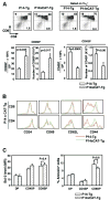
Expression of β-catenin increases CD8SP thymocytes in P14-Tg mice. A, Thymocyte development in P14-Tg and P14 × CAT-Tg mice analyzed by flow cytometry. Thymocytes from control P14-Tg mice or P14 × CAT-Tg were stained with Abs to CD4, CD8, and clonotypic Vα2 TCR and analyzed, as described in Fig. 2A. Left two bar graphs show percentage of CD8SP thymocytes (relative to DP thymocytes) and absolute number of CD8SP thymocytes; right two bar graphs show those of CD4SP thymocytes (n = 8). B, Phenotype of mature thymocytes. Expression of surface markers on CD8 and CD4SP thymocytes from P14 × CAT-Tg or control P14-Tg mice was analyzed by flow cytometry (n = 6). C, Bcl-2 expression and apoptosis in thymocyte subpopulations. Left panel, Intracellular Bcl-2 level in thymocyte subpopulations from P14 × CAT-Tg or control P14-Tg mice was shown (n = 3); right panel, percentage of annexin V+ cells in thymocyte subpopulations from P14 × CAT-Tg or control P14-Tg mice was shown (n = 3).
FIGURE 4
FIGURE 4
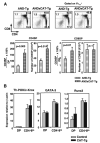
Expression of β-catenin does not alter lineage commitment. A, Thymocyte development in AND-Tg and AND × CAT-Tg mice analyzed by flow cytometry. Thymocytes were stained with Abs to CD4, CD8, and clonotypic Vα11 TCR. Left two bar graphs show percentage of CD4SP thymocytes (relative to DP thymocytes) and absolute number of CD4SP thymocytes; right two bar graphs show those of CD8SP thymocytes (n = 6). B, Expression of genes involved in lineage commitment. Expression of Th-POK, GATA-3, and Runx3 genes in sorted DP and CD4+8low thymocytes from AND × CAT-Tg or control AND-Tg mice was analyzed by real-time RT-PCR. Relative expression of the gene normalized to β-actin is shown (n = 3 independent experiments).
FIGURE 5
FIGURE 5
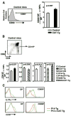
β-Catenin expression regulates positive selection intermediates. A, Histogram shows CD69 expression on DP thymocytes and the gate used to determine CD69+DP thymocytes from control mice. Graph shows percentage of CD69+DP thymocytes in control or CAT-Tg mice (n = 8 independent experiments). B, Dot plot shows the gate used to identify the CD4+8low population in control thymocytes. Bar graphs show percentage of CD4+8low thymocytes (relative to DP thymocytes) in CAT-Tg (n = 8), CAT-KO (n = 7), P14 × CAT-Tg (n = 8), and AND × CAT-Tg (n = 6) mice. C, Surface expression of IL-7Rα (upper panels) and CCR7 (lower panels) on DP and CD8SP thymocytes from control P14-Tg mice or P14 × CAT-Tg mice, analyzed by flow cytometry. Data are representative of four independent analyses.
FIGURE 6
FIGURE 6
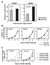
Expression of Tg β-catenin increases generation of mature thymocytes and accelerates maturation of CD8SP thymocytes. A, Maturation of CD4SP and CD8SP thymocytes in FTOCs. Embryonic day 17.5 fetal thymic lobes from control or CAT-Tg mice were placed in FTOCs. On day 3 of culture, thymocytes were harvested, treated with pronase (to remove surface CD4 and CD8), and then cultured overnight so that they would re-express CD4 and/or CD8 molecules that were being actively synthesized. Thymocytes were then analyzed by flow cytometry, and numbers of CD4SP and TCRβhigh CD8SP thymocytes per thymic lobe are shown (n = 3). B, Rate of generation of mature thymocytes studied using long-term BrdU labeling. Control and CAT-Tg mice were injected with BrdU and then maintained on BrdU-containing drinking water for 4 days. On each day, thymocytes from three mice of each genotype were stained with Abs to CD4, CD8, CD24, and BrdU, and analyzed by flow cytometry. Shown are BrdU+ cells in each thymocyte subpopulation as a percentage of the total determined each day after initial injection. C, Kinetics of CD4SP and CD8SP thymocyte generation. In the 4-day BrdU-labeling assay, the number of BrdU+ CD4SP or CD8SP thymocytes in control or CAT-Tg mice was determined each day after initial BrdU injection.

"VSports最新版本" References

    1. Jameson SC, Hogquist KA, Bevan MJ. Positive selection of thymocytes. Annu Rev Immunol. 1995;13:93–126. - PubMed
    1. Fowlkes BJ, Schweighoffer E. Positive selection of T cells. Curr Opin Immunol. 1995;7:188–195. - PubMed
    1. Bosselut R. CD4/CD8-lineage differentiation in the thymus: from nuclear effectors to membrane signals. Nat Rev Immunol. 2004;4:529–540. - V体育平台登录 - PubMed
    1. Germain RN. T-cell development and the CD4-CD8 lineage decision. Nat Rev Immunol. 2002;2:309–322. - PubMed
    1. Singer A, Bosselut R. CD4/CD8 coreceptors in thymocyte development, selection, and lineage commitment: analysis of the CD4/CD8 lineage decision. Adv Immunol. 2004;83:91–131. - "VSports手机版" PubMed

"V体育平台登录" Publication types

LinkOut - more resources