<kbd date-time="3UpvY2"></kbd><del id="kyTUN"></del> Skip to main page content
U.S. flag

An official website of the United States government

Dot gov

The . gov means it’s official. Federal government websites often end in . gov or . mil. Before sharing sensitive information, make sure you’re on a federal government site. VSports app下载.

Https

The site is secure. The https:// ensures that you are connecting to the official website and that any information you provide is encrypted and transmitted securely V体育官网. .

. 2007 Jun;18(6):2002-12.
doi: 10.1091/mbc.e06-09-0830. Epub 2007 Mar 14.

Superoxide flux in endothelial cells via the chloride channel-3 mediates intracellular signaling

Affiliations

Superoxide flux in endothelial cells via the chloride channel-3 mediates intracellular signaling (V体育ios版)

Brian J Hawkins et al. Mol Biol Cell. 2007 Jun.

VSports在线直播 - Abstract

Reactive oxygen species (ROS) have been implicated in both cell signaling and pathology. A major source of ROS in endothelial cells is NADPH oxidase, which generates superoxide (O(2)(. -)) on the extracellular side of the plasma membrane but can result in intracellular signaling. To study possible transmembrane flux of O(2)(. -), pulmonary microvascular endothelial cells were preloaded with the O(2)(. -)-sensitive fluorophore hydroethidine (HE). Application of an extracellular bolus of O(2)(. -) resulted in rapid and concentration-dependent transient HE oxidation that was followed by a progressive and nonreversible increase in nuclear HE fluorescence. These fluorescence changes were inhibited by superoxide dismutase (SOD), the anion channel blocker DIDS, and selective silencing of the chloride channel-3 (ClC-3) by treatment with siRNA VSports手机版. Extracellular O(2)(. -) triggered Ca(2+) release in turn triggered mitochondrial membrane potential alterations that were followed by mitochondrial O(2)(. -) production and cellular apoptosis. These "signaling" effects of O(2)(. -) were prevented by DIDS treatment, by depletion of intracellular Ca(2+) stores with thapsigargin and by chelation of intracellular Ca(2+). This study demonstrates that O(2)(. -) flux across the endothelial cell plasma membrane occurs through ClC-3 channels and induces intracellular Ca(2+) release, which activates mitochondrial O(2)(. -) generation. .


Figures
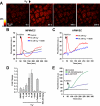
Figure 1.
Figure 1.
Hydroethidine (HE) oxidation with addition of extracellular O2.−. (A) A single bolus of O2.− was delivered to HE-loaded MPMVECs and recorded every 5 s. (B) Tracing indicating mean nuclear fluorescence of all cells in the field after addition of DMSO vehicle (control) or O2.− with or without preincubation with DIDS (200 μM). (C) Same experiment as B using HPMVECs. (D) Peak fluorescence change in MPMVECs (fold change normalized to baseline) in response to DMSO (vehicle control, n = 5), O2.− at 0.5 μM (n = 5), 2 μM (n = 5), 5 μM (n = 4), and 10 μM (n = 5), and H2O2 (500 μM; n = 3). The effects of SOD (2500 U/ml; n = 5), catalase (1000 U/ml; n = 3), and DIDS (200 μM; n = 5) were evaluated in the presence of 10 μM O2.−. (E) Mean cellular nuclear HE fluorescence after treatment with mitochondrial complex III inhibitor antimycin A (20 μM; n = 3) or the uncoupler FCCP (2 μM; n = 3).
Figure 2.
Figure 2.
HE oxidation transient in a cell-free system. (A) Fluorescence change was measured in a fluorimeter after addition of agent to PBS containing 40 μM HE. Additions were DMSO vehicle, KO2 (200 μM), xanthine/xanthine oxidase (X/XO; 50 mU), H2O2 (5 mM), KOH (200 μM), and KO2 predismutated into H2O2 with SOD. (B) Quantitation of the normalized peak and postpeak HE fluorescence increase over baseline after addition of varying concentrations of KO2. Linear regression lines were calculated from the mean values of three independent experiments.
Figure 3.
Figure 3.
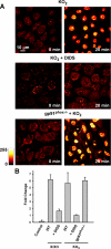
The stable increase of nuclear HE fluorescence by extracellular O2.− is blocked by DIDS but is gp91phox independent. (A) HE fluorescence in MPMVECs before and 20 min after exposure to extracellular O2.− (10 μM KO2) and in the absence (top panels) and presence of DIDS (200 μM). (B) HE fluorescence expressed as fold change versus baseline (zero time) was measured in untreated MPMVECs at 20 min after addition of O2.− (10 μM KO2 and 20 mU X/XO) with or without preincubation with DIDS (200 μM) and in gp91phox null cells. Control represents no additions. Results are mean ± SE; n = 3.
Figure 4.
Figure 4.
Receptor-mediated endothelial cell O2.− generation results in a stable increase of nuclear HE fluorescence. HPMVECs were loaded with HE (10 μM) and stimulated with Ang II (2 μM) or thrombin (0.5 U/ml) for 1 h with or without pretreatment with the NADPH oxidase inhibitor apocynin (Apo; 2 μM) or the anion channel blocker DIDS (300 μM). Nuclear HE fluorescence was quantitated from the confocal microscopic images. Data represent mean ± SE of five independent fields (n = 3).
Figure 5.
Figure 5.
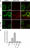
Mitochondrial O2.− production in response to extracellular O2.−. (A) Images of MitoGFP-transfected MPMVECs incubated with the mitochondrial O2.−-sensitive fluorescent dye MitoSox Red (1.25 μM) after extracellular bolus of O2.− with or without DIDS (200 μM). Images were taken 20 min after KO2 addition. All cells in the field show increased MitoSox Red fluorescence with addition of O2.−. Cells where MitoGFP is expressed show colocalization with MitoSox Red (merge panels). (B) Quantitation of MitoSox Red fluorescence at 20 min expressed as fold change versus baseline (zero time) for untreated cells (control) or cells treated with Tg (2 μM) or O2.− (10 μM) with or without DIDS (200 μM) or BAPTA (50 μM). Results are mean ± SE; n = 3.
Figure 6.
Figure 6.
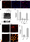
ClC-3 knockdown attenuates intracellular HE oxidation. (A) Electroporation of siRNA effectively delivers siRNA to MPMVECs. Images represent cy3-labeled GAPDH siRNA counterstained with the nuclear marker DAPI. (B) CIC-3, CIC-4, and β-actin mRNA expression in MPMVECs 60 h after transfection with one of two different CIC-3 sequences or negative control siRNA (250 pmol). Bottom, Western blot of ClC-3 protein expression in wild-type (WT) MPMVECs and at 72 h after transfection with ClC-3 #1 or negative control siRNA (control). (C) HE fluorescence transient in WT (n = 5), ClC-3 #1 (n = 5) and #2 (n = 5) and negative control (n = 5) siRNA transfected MPMVECs after addition of 10 μM KO2 normalized to fold change versus baseline. (D) HE fluorescence at baseline and at 20 min after exposure to extracellular O2.− (10 μM) in ClC-3 siRNA #1 and negative control siRNA-transfected MPMVECs. (E) Quantitation of HE fluorescence normalized to fold change versus baseline for control and ClC-3 #1-treated MPMVECs (n = 3).
Figure 7.
Figure 7.
Effect of extracellular O2.− on ΔΨm. (A) Time-lapse images of MPMVECs loaded with the mitochondrial potentiometric dye rhodamine 123 (25 μM) before and after addition of KO2 (10 μM). (B) Representative tracing of nuclear rhodamine 123 fluorescence after addition of DMSO and KO2 (10 μM) with or without DIDS (200 μM). (C) Representative tracing of the cytosolic Ca2+ indicator dye Fluo4 after application of KO2 (10 μM) in the absence and presence of the Ca2+ chelator BAPTA (50 μM) normalized to baseline fluorescence. (D) Representative tracing of nuclear rhodamine 123 fluorescence after addition of DMSO and KO2 (10 μM) with or without pretreatment with thapsigargin (Tg; 2 μM). Dissipation of ΔΨm is demonstrated by addition of the mitochondrial uncoupler FCCP (2 μM). (D) Representative tracings of rhodamine 123 fluorescence in HPMVECs after addition of KO2 (5 μM) or KCl (20 mM). Representative tracings are indicative of three independent experiments.
Figure 8.
Figure 8.
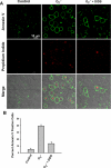
Extracellular O2.− leads to apoptosis in MPMVECs. MPMVECs were grown on coverslips and subjected to an extracellular bolus of KO2 (10 μM) with or without DIDS pretreatment (200 μM). (A) Cells were stained 3 h post-O2.− exposure for annexin V and propidium iodide. (B) The percentage of annexin V–positive cells was determined for 10 fields in each of 3 independent experiments.

References

    1. Aon M. A., Cortassa S., Marban E., O'Rourke B. Synchronized whole cell oscillations in mitochondrial metabolism triggered by a local release of reactive oxygen species in cardiac myocytes. J. Biol. Chem. 2003;278:44735–44744. - PubMed (VSports注册入口)
    1. Babior B. M. NADPH oxidase: an update. Blood. 1999;93:1464–1476. - PubMed
    1. Becker L. B., vanden Hoek T. L., Shao Z. H., Li C. Q., Schumacker P. T. Generation of superoxide in cardiomyocytes during ischemia before reperfusion. Am. J. Physiol. 1999;277:H2240–H2246. - PubMed
    1. Devadas S., Zaritskaya L., Rhee S. G., Oberley L., Williams M. S. Discrete generation of superoxide and hydrogen peroxide by T cell receptor stimulation: selective regulation of mitogen-activated protein kinase activation and fas ligand expression. J. Exp. Med. 2002;195:59–70. - "V体育官网" PMC - PubMed
    1. Fink B., Laude K., McCann L., Doughan A., Harrison D. G., Dikalov S. Detection of intracellular superoxide formation in endothelial cells and intact tissues using dihydroethidium and an HPLC-based assay. Am. J. Physiol. Cell Physiol. 2004;287:C895–C902. - "V体育2025版" PubMed

Publication types

MeSH terms