Skip to main page content (VSports app下载)
U.S. flag

An official website of the United States government

Dot gov

The . gov means it’s official. Federal government websites often end in VSports app下载. gov or . mil. Before sharing sensitive information, make sure you’re on a federal government site. .

Https

The site is secure V体育官网. The https:// ensures that you are connecting to the official website and that any information you provide is encrypted and transmitted securely. .

. 2007 Mar 13;104(11):4553-8.
doi: 10.1073/pnas.0610019104. Epub 2007 Mar 2.

Competition for antigen determines the stability of T cell-dendritic cell interactions during clonal expansion

Affiliations

VSports注册入口 - Competition for antigen determines the stability of T cell-dendritic cell interactions during clonal expansion

V体育官网 - Zacarias Garcia et al. Proc Natl Acad Sci U S A. .

Abstract

The regulation of T cell-dendritic cell (DC) contacts during clonal expansion is poorly defined. Although optimal CD4 T cell responses require prolonged exposure to antigen (Ag), it is believed that stable T cell-DC interactions occur only during the first day of the activation process. Here we show that recently activated CD4 T cells are in fact fully competent for establishing contact with Ag-bearing DC. Using two-photon imaging, we found that whereas prolonged interactions between activated T cells and Ag-bearing DCs were infrequent at high T cell precursor frequency, they were readily observed for a period of at least 2 days when lower numbers of T cells were used. We provide evidence that, when present in high numbers, Ag-specific T cells still gained access to the DC surface but were competing for the limited number of sites on DCs with sufficient peptide-MHC complexes for the establishment of a long-lived interaction. Consistent with these findings, we showed that restoration of peptide-MHC level on DCs at late time points was sufficient to recover interactions between activated T cells and DCs. Thus, the period during which CD4 T cells continue to establish stable interactions with DCs is longer than previously thought, and its duration is dictated by both Ag levels and T cell numbers, providing a feedback mechanism for the termination of CD4 T cell responses. VSports手机版.


Conflict of interest statement

The authors declare no conflict of interest.

Figures
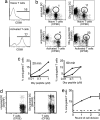
Fig. 1.
Fig. 1.
Activated T cells are fully competent for establishing conjugates with Ag-bearing DCs in vitro. (a) Naïve Marilyn CD4 T cells were activated in vitro by using anti-CD3/CD28 beads. After 48 h, T cells were stained for CD69. (b and c) Naïve or activated Marilyn CD4 T cells were labeled with 1 μM CFSE and incubated with DCs pulsed with the Dby peptide or left unpulsed and labeled with 1 μM SNARF. (b) The percentage of naïve or activated T cells conjugated to a DC was quantified by flow cytometry at 20 min. DCs were pulsed with 1 μM Dby peptide. (c) The percentage of conjugated naïve (open circles) or activated (filled circles) T cells was measured at 20 and 60 min for different peptide concentrations used to pulse DCs. (d) Naïve Marilyn T cells were labeled with CFSE and stimulated in vitro with anti-CD3/CD28 beads. After 48 h, activated T cells were incubated with an excess of SNARF-labeled DCs that were either left unpulsed (Left) or pulsed with the Dby peptide (Right), and cell conjugation was assessed after 60 min. (e) The graph shows the percentage of conjugated T cells among those T cells that have undergone the indicated number of cell division. Cell conjugation was performed by using Dby-pulsed DCs (solid line) or unpulsed DCs (dashed line). Values are means ± SD. Results are representative of three independent experiments.
Fig. 2.
Fig. 2.
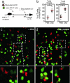
Activated T cells interact with Ag-bearing DCs in vivo. (a) Experimental setup. MHC class II-deficient mice were injected in the footpad with 2 × 10⁶ unpulsed wild-type female DCs labeled with a mixture of CFSE and SNARF dyes (0.5 μM and 1 μM, respectively). Recipients were also adoptively transferred with a 1:1 mixture of SNARF-labeled naïve Marilyn T cells and CFSE-labeled activated Marilyn T cells. At 24 h, each recipient received an i.v. injection of Dby peptide (50 μg) to specifically pulse the injected DCs present in the draining lymph node. After 30 min, popliteal lymph nodes were harvested, sectioned, and analyzed by confocal microscopy. (b) The percentage of activated or naïve T cells contacting a DC was measured on confocal images of lymph node sections. Each dot represents the value derived from an individual section. Mean values are indicated by a red bar. ∗, P = 0.06; ∗∗, P < 0.001. Results are representative of two independent experiments. (c and d) Confocal images of lymph node sections 30 min after injection of PBS (c) or Dby peptide (d). DCs are yellow, naïve T cells are red, and activated T cells are green. (Scale bar: 20 μm.)
Fig. 3.
Fig. 3.
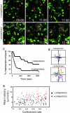
T cell precursor frequency dictates the efficiency of stable T cell–DC formation. B6 recipient mice were immunized by footpad injection of 1.5 × 10⁶ DCs derived from Ubi-GFP mice and pulsed with 100 nM Dby peptide. After 2–4 h, recipients were adoptively transferred with 1 × 10⁶ SNARF-labeled naïve Marilyn CD4 T cells alone or together with 9 × 10⁶ unlabeled Marilyn CD4 T cells (competitors). At 48 h after DC injection, intact popliteal lymph nodes were subjected to two-photon real-time imaging. (a and b) Time-frame images showing that long-lived T cell–DC interactions (marked by white circles) at 48 h occur in the absence (b) but not in the presence (a) of competitors. (c) Individual T cell–DC contacts (n = 35) were monitored over time, and the percentage of T cell–DC contacts maintained was graphed as a function of time. Long-lived interactions were observed only in the absence of competitors. (d) Individual T cell trajectories were analyzed in the absence (n = 50) or in the presence (n = 48) of competitors and graphed from a common origin. (e) T cells displayed higher velocities and confinement ratios in the presence of competitors. Individual CD4 T cells were tracked over time, and their mean velocity was graphed against their confinement ratio. Low confinement ratios reflect constrained movement. A total of 77 and 104 T cells tracks were analyzed in the presence and in the absence of competitors, respectively. Results are representative of at least four time-lapse movies obtained in two independent experiments.
Fig. 4.
Fig. 4.
T cell competition is not due to steric hindrance but to the lack of sites on DCs allowing the formation of stable interactions. Recipient mice were immunized by footpad injection of 1.5 × 10⁶ SNARF-labeled DCs pulsed with 100 nM Dby peptide. After 2–4 h, recipients were adoptively transferred with 1 × 10⁶ or 10 × 106 CFSE-labeled naïve Marilyn CD4 T cells. Intact popliteal lymph nodes were subjected to two-photon real-time imaging at 48 h after DC injection. (a) Representative images showing the density of CD4 T cells 48 h after transfer of 10 × 106 T cells. (b and c) Stable interactions but not transient encounters are limiting in the presence of high T cell numbers. Individual DCs were analyzed from two-photon time-lapse movies, and the number of transient encounters (<5 min) was recorded (over a fixed period of 25 min) as well as the number of stable interactions (>20 min). Mean values are indicated by a red bar. ∗, P < 0.001; ns, not significant (P = 0.32). Results are representative of at least three time-lapse movies obtained in two independent experiments.
Fig. 5.
Fig. 5.
T cell activation is reduced in the presence of high numbers of T cells. Recipient mice were immunized by footpad injection of 1.5 × 10⁶ SNARF-labeled DCs pulsed with either 100 nM (a) or 10 nM (b) Dby peptide. After 2–4 h, recipients were adoptively transferred with 1 × 10⁶ or 10 × 106 CFSE-labeled naïve Marilyn CD4 T cells. At various time points, popliteal lymph nodes were harvested and lymph node cells were analyzed by flow cytometry. Data are gated on CD4+CD45.1+ cells. At high T cell precursor frequency, T cell activation and proliferation were markedly reduced on a per-cell basis. Results are representative of three independent experiments.
Fig. 6.
Fig. 6.
Recovery of late T cell–DC interactions upon restoration of pMHC on DCs. (a) Experimental setup. MHC class II-deficient mice were injected in the footpad with DCs pulsed with 100 nM Dby peptide. After 2–4 h, recipients were adoptively transferred with 10 × 106 CFSE-labeled naïve Marilyn T cells. Recipient mice were injected i.v. with 50 μg of Dby peptide at 48 h after DC injection to restore pMHC levels at the surface of the transferred DCs. Control animals were injected with PBS. T cell–DC contacts were quantified 3 h after peptide (or PBS) injection. (b) The fraction of Marilyn CD4 T cells found to interact with a transferred DC after peptide or PBS injection was quantified on lymph node sections. Each dot represents the value derived from an individual confocal image of a lymph node section. The corresponding value was calculated for those T cells displaying the hallmark of T cell activation (large cells with dim CFSE intensity). Mean values are indicated by a red bar. (c and d) Representative images of lymph node section after PBS (c) or Dby (d) injection. ∗, P < 0.001. Results are representative of two independent experiments.

References

    1. Breart B, Bousso P. Curr Opin Immunol. 2006;18:483–490. - PubMed
    1. Mempel TR, Henrickson SE, Von Andrian UH. Nature. 2004;427:154–159. - PubMed
    1. Miller MJ, Safrina O, Parker I, Cahalan MD. J Exp Med. 2004;200:847–856. - PMC - PubMed
    1. Hugues S, Fetler L, Bonifaz L, Helft J, Amblard F, Amigorena S. Nat Immunol. 2004;5:1235–1242. - PubMed
    1. Zinselmeyer BH, Dempster J, Gurney AM, Wokosin D, Miller M, Ho H, Millington OR, Smith KM, Rush CM, Parker I, et al. J Exp Med. 2005;201:1815–1823. - V体育2025版 - PMC - PubMed

Publication types

MeSH terms (VSports注册入口)