Skip to main page content
U.S. flag

An official website of the United States government

Dot gov

The VSports app下载. gov means it’s official. Federal government websites often end in . gov or . mil. Before sharing sensitive information, make sure you’re on a federal government site. .

Https

The site is secure. The https:// ensures that you are connecting to the official website and that any information you provide is encrypted and transmitted securely. V体育官网.

. 2007 Mar;170(3):1077-85.
doi: 10.2353/ajpath.2007.060773.

Orthotopic microinjection of human colon cancer cells in nude mice induces tumor foci in all clinically relevant metastatic sites (V体育平台登录)

Affiliations

Orthotopic microinjection of human colon cancer cells in nude mice induces tumor foci in all clinically relevant metastatic sites

"VSports最新版本" María Virtudes Céspedes et al. Am J Pathol. 2007 Mar.

Abstract (V体育平台登录)

Despite metastasis as an important cause of death in colorectal cancer patients, current animal models of this disease are scarcely metastatic. We evaluated whether direct orthotopic cell microinjection, between the mucosa and the muscularis layers of the cecal wall of nude mice, drives tumor foci to the most relevant metastatic sites observed in humans and/or improves its yield as compared with previous methods. We injected eight animals each tested human colorectal cancer cell line (HCT-116, SW-620, and DLD-1), using a especially designed micropipette under binocular guidance, and evaluated the take rate, local growth, pattern and rate of dissemination, and survival time. Take rates were in the 75 to 88% range. Tumors showed varying degrees of mesenteric and retroperitoneal lymphatic foci (57 to 100%), hematogenous dissemination to liver (29 to 67%) and lung (29 to 100%), and peritoneal carcinomatosis (29 to 100%). Tumor staging closely correlated with animal survival. Therefore, the orthotopic cell microinjection procedure induces tumor foci in the most clinically relevant metastatic sites: colon-draining lymphatics, liver, lung, and peritoneum. The replication of the clinical pattern of dissemination makes it a good model for advanced colorectal cancer. Moreover, this procedure also enhances the rates of hematogenous and lymphatic dissemination at relevant sites, as compared with previously described methods that only partially reproduce this pattern VSports手机版. .


Figures
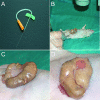
Figure 1
Figure 1
OCMI into the cecum of immunosuppressed mice. A: View of the micropipette made from Vitrex capillaries with a 250-μm-diameter tip. B: The cecum of anesthetized nude mice is exteriorized through a laparotomy. C: Two million human colorectal cancer cells, per animal, are injected, with a 30° angle under a binocular lens (×3). D: Reddish area depicts the tissue where tumor cell suspension has been injected.
Figure 2
Figure 2
Site of injection and pattern of tumor growth and invasion. A: The site of injection, 1 week after the OCMI procedure, showed the tumor cells located between the mucosa and the muscularis externa layers of the cecal wall. B: Tumors grow, both tangentially and circumferentially, and protrude into the lumen of the large bowel obstructing it. C–E: Tumors derived from HCT-116 (C), SW-620 (D), or DLD-1 (E) cell lines are poorly differentiated adenocarcinomas with high cellularity, focal areas of necrosis, frequent mitoses, and highly atypical nuclei, showing invasion of all of the cecal layers. F: Tumor cells infiltrate the lymphatics of the cecal wall (white asterisk). A, C–F: H&E stains.
Figure 3
Figure 3
Lymphatic, hematogenous, and peritoneal tumor spread in nude mice bearing tumors derived from OCMI-implanted cell lines A: Out of the local tumor (black asterisk), tumor cells spread to the mesenteric lymph nodes (white arrow). B: Microscopically, tumor foci in the mesenteric lymph nodes showed a rim of lymphocytes (white asterisk) compressed against the capsule by tumor cells (black asterisk). C: Representative foci of hematogenous dissemination to the liver, observed in the HCT-116 cell line. D: Tumor cells at the lung were found inside peribronchial vessels. E: Tumor cells invading the muscle of the diaphragm from its peritoneal surface. B–E: H&E stains.
Figure 4
Figure 4
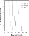
Survival curves of animals from the three studied groups. Survival time was the shortest in HCT-116 tumor-bearing mice, intermediate in SW-620 animals, and the longest in DLD-1 mice. Cumulative survival values were estimated using the Kaplan and Meier method and plotted versus time after injection. The log-rank test showed statistically significant differences in survival among groups (see also Table 2).

References

    1. Jemal A, Siegel R, Ward E, Murray T, Xu J, Smigal C, Thun MJ. Cancer statistics, 2006. CA Cancer J Clin. 2006;56:106–130. - PubMed
    1. Corpet DE, Pierre F. Point: from animal models to prevention of colon cancer. Systematic review of chemoprevention in min mice and choice of the model system. Cancer Epidemiol Biomarkers Prev. 2003;12:391–400. - PMC (VSports注册入口) - PubMed
    1. Donehower LA, Harvey M, Slagle BL, McArthur MJ, Montgomery CA, Jr, Butel JS, Bradley A. Mice deficient for p53 are developmentally normal but susceptible to spontaneous tumours. Nature. 1992;356:215–221. - PubMed
    1. Hoffman RM. Orthotopic metastatic mouse models for anticancer drug discovery and evaluation: a bridge to the clinic. Invest New Drugs. 1999;17:343–359. - PubMed
    1. Fu XY, Besterman JM, Monosov A, Hoffman RM. Models of human metastatic colon cancer in nude mice orthotopically constructed by using histologically intact patient specimens. Proc Natl Acad Sci USA. 1991;88:9345–9349. - PMC - PubMed

V体育ios版 - Publication types