Skip to main page content (VSports)
U.S. flag

An official website of the United States government

Dot gov

The . gov means it’s official. Federal government websites often end in . gov or VSports app下载. mil. Before sharing sensitive information, make sure you’re on a federal government site. .

Https

The site is secure. The https:// ensures that you are connecting to the official website and that any information you provide is encrypted and transmitted securely. V体育官网.

. 2007 Mar 7;26(5):1292-302.
doi: 10.1038/sj.emboj.7601586. Epub 2007 Feb 15.

Chromatin remodeling by the SWI/SNF-like BAF complex and STAT4 activation synergistically induce IL-12Rbeta2 expression during human Th1 cell differentiation

Affiliations

"V体育官网入口" Chromatin remodeling by the SWI/SNF-like BAF complex and STAT4 activation synergistically induce IL-12Rbeta2 expression during human Th1 cell differentiation

"VSports手机版" Fabrice A Letimier et al. EMBO J. .

Abstract

Interleukin-12 (IL-12) is a key cytokine for the development of T helper type 1 (Th1) responses; however, naïve CD4(+) T cells do not express IL-12Rbeta2, and are therefore unresponsive to IL-12. We have examined the mechanisms that control Th1-specific expression of the human IL-12Rbeta2 gene at early time points after T-cell stimulation VSports手机版. We have identified a Th1-specific enhancer element that binds signal transducer and activator of transcription 4 (STAT4) in vivo in developing Th1 but not Th2 cells. T-cell receptor (TCR) signaling induced histone hyperacetylation and recruitment of BRG1, the ATPase subunit of the SWI/SNF-like BAF chromatin remodeling complex, to the IL-12Rbeta2 regulatory regions and was associated with low-level gene transcription at the IL-12Rbeta2 locus. However, high-level IL-12Rbeta2 expression required TCR triggering in the presence of IL-12. Our results indicate a synergistic role of TCR-induced chromatin remodeling and cytokine-induced STAT4 activation to direct IL-12Rbeta2 expression during Th1 cell development. .


VSports手机版 - Figures
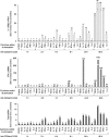
Figure 1
Figure 1
Cytokines rapidly induce IL-12Rβ2, IFN-γ, and T-bet transcripts following stimulation of naïve human CD4+ T cells. Naïve CD4+ T cells were purified from cord blood and stimulated with anti-CD3/CD28 antibodies in the presence of the indicated cytokines. Cells were harvested at the indicated times and the mRNA levels of IL-12Rβ2, IFN-γ, and T-bet were determined by kinetic real-time PCR. The fold increase of IL-12Rβ2 (upper panel), IFN-γ (middle panel), and T-bet transcripts (lower panel) when comparing stimulated T cells to naïve CD4+ T cells is shown.
Figure 2
Figure 2
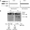
(A) The IL-12Rβ2 promoter is active in lymphoid and non-lymphoid cells. A 740 bp fragment (−173 to +572) of the IL-12Rβ2 gene was cloned in both directions upstream of the luciferase gene. These constructs were transiently transfected into Jurkat cells (left panel) or COS7 cells (right panel) and luciferase activity in cell lysates was measured 24 h after transfection. Transfection efficiency was normalized by measuring β-galactosidase activity of a CMV-βgal vector cotransfected in the cells. Results are representative of five independent experiments. (B) Mapping of DNAse I-hypersensitive sites (HS) in the IL-12Rβ2 gene in Th1 and Th2 cells. Nuclei were purified from Th1 (right panel) or Th2 (left panel) cells 6 days after priming and treated with increasing amounts of DNase I (lanes 2–5 and 7–10). Genomic DNA was digested with PvuII and Southern blot analysis performed with a 0.5 kb PvuII–BamHI genomic fragment as a probe. The lower panel indicates the positions of HS with respect to the PvuII restriction site, the GAS elements, the mRNA start site, and the probe used for Southern blot analysis.
Figure 3
Figure 3
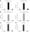
Chromatin remodeling at the IL-12Rβ2 enhancer and promoter is restricted to the Th1 subset. Histone 4 (H4) acetylation (A) and histone 3 lysine 4 (H3 K4) trimethylation (B) were analyzed in naïve CD4+ T cells and in Th1 and Th2 cells at day 5 after stimulation. Quantification of the specific enrichment of IL-12Rβ2 enhancer or promoter fragments following ChIP with anti-acetyl H4, anti-trimethyl H3 K4 was performed by Taqman real-time PCR. The mRNA levels of IL-12Rβ2 (C) and IFN-γ (D) were determined in the same samples by real-time PCR with Taqman probes and calculated relative to the expression of the target gene in naïve T cells.
Figure 4
Figure 4
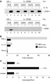
(A) STAT4 binds to GAS elements in the IL-12Rβ2 regulatory region. Biotinylated oligonucleotide probes encompassing the four GAS elements from the IL-12Rβ2 gene (lanes 3–10) or the GAS element present in the human IRF-1 promoter (lanes 1 and 2) were incubated with nuclear extracts from IL-12-treated or untreated Th1 cells (upper panel), or IFN-γ-treated or untreated Th2 cells (lower panel). Proteins bound to the probes were precipitated with streptavidin–agarose, separated on SDS gels, and transferred to PVDF membranes. The membranes were probed with antibodies to STAT4 (upper panel) or STAT1 (lower panel). (B) IL-12Rβ2 GAS elements mediate IL-12-induced transcriptional activation. The beta2-Luc construct (black bars) containing all four IL-12Rβ2 GAS elements was compared with the IRF1-Luc construct (gray bars) for responsiveness to IL-12- and IFN-γ mediated transcriptional activation. Jurkat cells were transiently cotransfected as described in Supplementary data section to reconstitute IL-12 signaling plus the reporter construct. Cells were left untreated or stimulated for 20 h with IL-12 or IFN-γ. Luciferase activity was measured 24 h after transfection. Results were normalized by measuring the activity of a cotransfected CMV-βgal vector. Transfections were performed in triplicate and the experiment is representative of three. (C) IL-12Rβ2 GAS elements mediate Th1-specific expression of a reporter gene. Naïve CD4+ T cells were transfected with the beta2-Luc construct containing the four GAS elements from the IL-12Rβ2 gene. Cells were left unstimulated or stimulated in Th1 or Th2 conditions. Luciferase activity was measured 24 h after stimulation and normalized as above. The bars represent mean and s.d. from one representative experiment out of two.
Figure 5
Figure 5
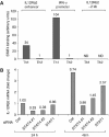
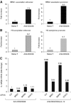
(A) STAT4 is bound to the enhancer of the IL-12Rβ2 gene in vivo. Naïve CD4+ T cells were stimulated for 20 h with anti-CD3/CD28 in Th1 or Th2 conditions. ChIP assays were performed with anti-STAT4 antibodies or rabbit IgG as control (see Materials and methods). The enrichment of IL-12Rβ2 enhancer sequences in the precipitate was measured by real-time PCR using Taqman probes. Shown is binding of STAT4 to the IL-12Rβ2 enhancer and the IFN-γ promoter in Th1 relative to Th2 cells. No STAT4 binding to a fragment spanning GAS elements 3 and 4 (a sequence element 2 kb upstream of the IL-12Rβ2 mRNA start site) was detected (ND, amplification equal to background). (B) Knock-down of STAT4 decreases IL-12Rβ2 expression. Naïve CD4+ T cells were transfected with a control siRNA oligo (CTRL), siRNA oligos targeting STAT4 (STAT4 #1 and STAT4 #2), or an oligo targeting STAT1. Cells were stimulated with anti-CD3/CD28 in the presence of IL-12 and harvested after 24 or 48 h. Transcripts encoding IL-12Rβ2, STAT4, and STAT1 were measured by real-time PCR using Taqman probes. Shown are the expression levels of IL-12Rβ2 mRNA relative to the control at 24 h after stimulation. STAT4 and STAT1 mRNAs were inhibited 60–65 and 75–85%, respectively (not shown). Similar results were observed in five experiments.
Figure 6
Figure 6
IL-12Rβ2 enhancer and promoter undergo rapid epigenetic changes following T-cell stimulation. Naïve T-cells were left untreated or treated for 20 h with IFN-α (1000 U/ml) alone (naïve T). Alternatively, naïve T cells were stimulated with anti-CD3/CD28 antibodies for 20 h in the absence of exogenously added cytokines or in the presence of IFN-α (1000 U/ml), IL-12 (5 ng/ml), or IL-4 (1 ng/ml). Following ChIP of acetylated histone H3, IL-12Rβ2 enhancer fragments (A) or promoter fragments (B) were amplified by Taqman real-time PCR. The mRNA levels of IL-12Rβ2 (C), IFN-γ (D), T-bet (E), and GATA-3 (F) were determined in the same samples by real-time PCR with Taqman probes. Shown are the expression levels, normalized to 18S RNA amplification, in each sample and calculated relative to the expression of the target gene in naïve T cells.
Figure 7
Figure 7
(A) Stimulation of naïve T cells induces recruitment of the BAF chromatin remodeling complex to the IL-12Rβ2 locus. Naïve CD4+ T cells were harvested after purification or after stimulation for 20 h with anti-CD3/CD28. (A) ChIP assays were performed with anti-BRG1 or control antibodies, and the enrichment of IL-12Rβ2 enhancer or promoter sequences in the precipitate was measured by real-time PCR. Similar results were obtained using two different anti-BRG1 antibodies. (B) Histone H3 acetylation was analyzed at the IL-12Rβ2 enhancer and promoter in the same samples as described above. (C) Knock-down of BRG1 results in reduced IL-12Rβ2 expression. Naïve CD4+ T cells were transfected with a control siRNA oligo (CTRL) or siRNA oligos targeting BRG1, SNF2h, or STAT4. Cells were stimulated with anti-CD3/CD28 in the absence or presence of IL-12 and harvested after 24 h. Transcripts encoding IL-12Rβ2 were determined by real-time PCR. Expression levels of IL-12Rβ2 mRNA are shown relative to the control. BRG1, SNF2h, and STAT4 mRNAs were inhibited 70, 90–95, and 60–65%, respectively (not shown). Similar results were observed in three experiments.
Figure 8
Figure 8
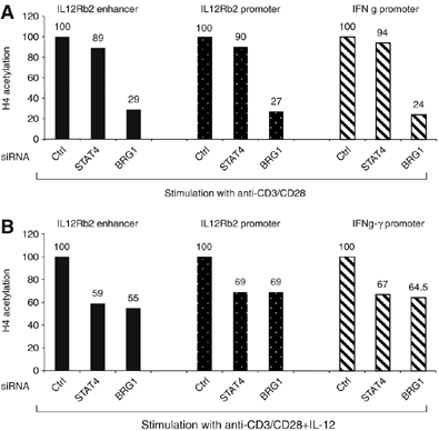
Downregulation of BRG1 and STAT4 inhibits H4 acetylation at the IL-12Rβ2 regulatory regions. Naïve T cells were transfected with the indicated siRNA oligos and stimulated for 24 h with anti-CD3/CD28 antibodies in the absence (A) or presence (B) of IL-12. ChIP assays were performed with anti-acetyl H4 or control antibodies and regions spanning the IL-12Rβ2 enhancer, promoter, or the IFN-γ promoter were amplified by real-time PCR. Values are expressed relative to the control siRNA sample.
Figure 9
Figure 9
Rapid recruitment of BRG1 and STAT4 to the IL-12Rβ2 regulatory elements during Th1 cell differentiation. Naïve T cells were stimulated with anti-CD3/CD28 antibodies+IL-12 for the indicated times. ChIP assays were performed with anti-BRG1 (A), anti-STAT4 (B), anti-acetyl-H4 (C), or control antibodies. Real-time PCR amplification of the enhancer and the promoter region are represented relative to the unstimulated samples. No specific amplification was observed in samples precipitated with control antibodies (not shown).
Figure 10
Figure 10
Induction of IL-12Rb2 expression during Th1 development: a model. A color version of this figure is available at The EMBO Journal Online (Supplementary Figure 5).

References

    1. Afkarian M, Sedy JR, Yang J, Jacobson NG, Cereb N, Yang SY, Murphy TL, Murphy KM (2002) T-bet is a STAT1-induced regulator of IL-12R expression in naive CD4+ T cells. Nat Immunol 3: 549–557 - PubMed
    1. Agarwal S, Rao A (1998) Modulation of chromatin structure regulates cytokine gene expression during T cell differentiation. Immunity 9: 765–775 - PubMed
    1. Ansel KM, Lee DU, Rao A (2003) An epigenetic view of helper T cell differentiation. Nat Immunol 4: 616–623 - PubMed
    1. Athie-Morales V, Smits HH, Cantrell DA, Hilkens CM (2004) Sustained IL-12 signaling is required for Th1 development. J Immunol 172: 61–69 - PubMed
    1. Avni O, Lee D, Macian F, Szabo SJ, Glimcher LH, Rao A (2002) T(H) cell differentiation is accompanied by dynamic changes in histone acetylation of cytokine genes. Nat Immunol 3: 643–651 - PubMed (V体育2025版)

VSports - Publication types

MeSH terms