Skip to main page content
U.S. flag

An official website of the United States government

Dot gov

The . gov means it’s official. Federal government websites often end in VSports app下载. gov or . mil. Before sharing sensitive information, make sure you’re on a federal government site. .

Https

The site is secure. The https:// ensures that you are connecting to the official website and that any information you provide is encrypted and transmitted securely V体育官网. .

. 2007 May 15;109(10):4336-42.
doi: 10.1182/blood-2006-09-046201. Epub 2007 Jan 23.

Expansion of spleen myeloid suppressor cells represses NK cell cytotoxicity in tumor-bearing host

Affiliations

Expansion of spleen myeloid suppressor cells represses NK cell cytotoxicity in tumor-bearing host (VSports)

Cunren Liu et al. Blood. .

Abstract

Tumor growth promotes the expansion of myeloid suppressor cells. An inverse correlation between natural killer (NK) cell activation and myeloid suppressor cell (MSC) expansion in tumor-bearing patients and mice prompted us to investigate the role of MSCs in controlling NK antitumor cytotocixity. After adoptive transfer to naive recipients, CD11b(+)Gr-1(+) MSCs freshly isolated from spleens of tumor-bearing mice but not naive mice were able to inhibit NK cell cytotoxicity. An in vivo imaging analysis indicates that the removal of tumors resulted in a significant increased ability (P < VSports手机版. 05) in NK cell cytotoxicity to eliminate injected YAC-1 cells from the lungs. Fluorescence-activated cell sorter (FACS) analysis of the composition of lung leukocytes further indicates that the removal of tumors also leads to the reduction of MSCs accumulated in the lung. These data suggest that MSCs suppress NK cell cytotoxicity. The inhibition of NK cell cytotoxicity is cell-cell contact dependent. Inhibition of perforin but not granzyme B production was responsible for MSC-mediated inhibition of NK cytotoxicity. Western blot analyses further suggests that MSCs suppress IL-2-mediated NK cell cytotoxicity by affecting the activity of Stat5. .


VSports app下载 - Figures

Figure 1
Figure 1
NK cell cytotoxicity is suppressed in tumor-bearing mice. Seven-week-old BALB/c mice were injected subcutaneously with TS/A tumor cells (3 × 10⁵) or PBS as a control. Four weeks after tumor challenge, the mice were anesthetized and injected intravenously with YAC-1–Luc cells (1 × 10⁶). After injection of D-luciferin the mice were imaged at hours 0, 2, 4, and 6 (A), and the total photon count per minute (photons per minute) calculated (5 animals) using Living Image software. The efficiency of NK cell killing of injected YAC-1–Luc cells was determined by measuring the numbers of photons collected at the imaging time points divided by at 0 hours collected (B). *P < .05; **P < .001. On completion of the imaging studies, NK cells (DX5+) were isolated from the spleen, and then stimulated with recombinant IL-2 (100 U/mL) for 5 days. After the incubation period, the NK cells were added to YAC-1–Luc or TS/A-Luc cells at varying effector-target (E/T) ratios (10:1, 20:1, and 40:1) as indicated in panels C and D. The cytotoxicity of NK cells to YAC-1–Luc (C) or TS/A-Luc (D) was determined using an NK cell cytotoxic assay as described in “Cytotoxicity assay.” The data represent the mean ± SEM from 5 mice from each group.
Figure 2
Figure 2
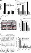
Spleen MSCs suppress NK cell cytotoxicity in vivo. Tumor-bearing mice at 4 weeks after tumor injection were killed and spleen cells were isolated. (A) The percentages of DX5, F4/80, CD11b, and Gr-1+ cells were determined by FACS analysis as described in “Flow cytometry analysis.” The data represent the mean ± SEM from 5 mice from each group. (B) CD11b+Gr-1+ cells were isolated from the spleens of 9-week-old TS/A tumor-bearing BALB/c mice or naive mice. Increased numbers (1 × 10⁶, 3 × 10⁶, and 6 × 10⁶) of sorted CD11b+Gr-1+ MSCs were transferred intravenously into 2-month-old BALB/c female mice (n = 5 per group). Twenty-four hours after adoptive transfer, the efficiency of NK cell killing of injected YAC-1–Luc cells was determined by measuring the numbers of photons collected at 6 hours divided by the photons collected at 0 hours. *P < .05; **P < .001. (C) Female BALB/c mice (n = 4): TS/A tumor-bearing mice with tumor debulked surgically (surgery removal), tumor-intact mice (nonremoval), and non–tumor-bearing PBS control mice (naive mice). Two weeks later, an in vivo measurement of NK cell cytotoxicity was determined by injection of YAC-1–Luc using an identical protocol, as described in Figure 1. The efficiency of NK cell killing of injected YAC-1–Luc cells was determined by measuring the numbers of photons collected at 2 hours and 4 hours divided by the photons collected at 0 hours. *P < .05; **P < .001. The data represent the mean ± SEM of 2 independent experiments (n = 4) (C, right panel). (D) After imaged mice were killed, the percentages of leukocytes in the lung were determined in the gated R1 region of a FACS analysis (top left). The presence of CD11b+Gr-1+, DX5+, CD8+, and CD11c+ cells was determined (left, representative plots). Results obtained from 2 independent experiments with replica 4 mice in each experiment were pooled and are presented as the mean ± SEM. *P < .05; **P < .01.
Figure 3
Figure 3
Spleen MSCs suppress NK cell cytotoxicity in a cell-cell contact–dependent manner. Splenic MSCs (CD11b+Gr-1+) from naive or tumor-bearing BALB/c mice were FACS sorted and cocultured with DX5+ NK cells at the ratios between 1:0 to 1:2 (NK/MSC) in a 24-well plate (A) or in a Transwell system (B). After 1 day (C) or 5 days (A-B) of NK-MSC coincubation, NK cell cytotoxicity against YAC-1–Luc cells (YAC-1–Luc/NK, 1:20) was assayed as described in “Cytotoxicity assay.” The data represent NK cell killing YAC-1–LUC activity at 4 hours after addition of YAC-1–Luc cells. Data are mean (± SEM) of triplicate wells of 3 independent experiments. *P < .05; **P < .001.
Figure 4
Figure 4
MSCs preferentially inhibit the production of perforin from NK cells. Spleen DX5+ NK cells were cocultured with spleen MSCs for 5 days at the ratio of 1:1 (A) or at varying ratios as indicated in panel B. DX5+ NK cells were then isolated with DX5 antibody-coated magnetic beads using the method as described previously. NK cells (1 × 10⁶) were lysed in protein lysis buffer, and 50 μg total protein from each lysate was resolved on a 10% SDS PAGE gel. The proteins were then transferred to a nitrocellulose membrane, and the blots were probed with the indicated antibodies. The data are representative of 3 independent experiments. β-actin served as an internal control to confirm equivalent protein loading.
Figure 5
Figure 5
Coculture of NK-MSCs results in the inhibition of phosphorylation of NK cell Stat5. Protein lysates were produced identically to those in Figure 4. Protein lysates from NK-MSCs cocultured at 1:1 and varying ratios (B) were run in the Western blot. The data are representative of 3 independent experiments. β-Actin was used as an internal control to confirm equivalent protein loading.

References (VSports app下载)

    1. Gallina G, Dolcetti L, Serafini P, et al. Tumors induce a subset of inflammatory monocytes with immunosuppressive activity on CD8+ T cells. J Clin Invest. 2006;116:2777–2790. - PMC - PubMed
    1. Frey AB. Myeloid suppressor cells regulate the adaptive immune response to cancer. J Clin Invest. 2006;116:2587–2590. - PMC - PubMed
    1. Kim R, Emi M, Tanabe K. Cancer immunosuppression and autoimmune disease: beyond immunosuppressive networks for tumour immunity. Immunology. 2006;119:254–264. - PMC - PubMed
    1. Valenti R, Huber V, Filipazzi P, et al. Human tumor-released microvesicles promote the differentiation of myeloid cells with transforming growth factor-beta-mediated suppressive activity on T lymphocytes. Cancer Res. 2006;66:9290–9298. - PubMed (V体育ios版)
    1. Bunt SK, Sinha P, Clements VK, Leips J, Ostrand-Rosenberg S. Inflammation induces myeloid-derived suppressor cells that facilitate tumor progression. J Immunol. 2006;176:284–290. - PubMed

Publication types

MeSH terms