<u date-time="T463nN"></u><sub date-time="cewdD"></sub><sub date-time="1zynl"><small dropzone="xP0jwkyM"></small></sub> Skip to main page content
U.S. flag

An official website of the United States government

Dot gov

The . gov means it’s official. Federal government websites often end in . gov or . mil. Before sharing sensitive information, make sure you’re on a federal government site VSports app下载. .

Https

The site is secure. The https:// ensures that you are connecting to the official website and that any information you provide is encrypted and transmitted securely V体育官网. .

. 2007 Jan;26(1):79-92.
doi: 10.1016/j.immuni.2006.10.018. Epub 2006 Dec 21.

Antigen-loading compartments for major histocompatibility complex class II molecules continuously receive input from autophagosomes (VSports手机版)

Affiliations

Antigen-loading compartments for major histocompatibility complex class II molecules continuously receive input from autophagosomes

Dorothee Schmid (VSports app下载) et al. Immunity. 2007 Jan.

Abstract

Major histocompatibility complex (MHC) class II molecules present products of lysosomal proteolysis to CD4(+) T cells. Although extracellular antigen uptake is considered to be the main source of MHC class II ligands, a few intracellular antigens have been described to gain access to MHC class II loading after macroautophagy. However, the general relevance and efficacy of this pathway is unknown VSports手机版. Here we demonstrated constitutive autophagosome formation in MHC class II-positive cells, including dendritic, B, and epithelial cells. The autophagosomes continuously fuse with multivesicular MHC class II-loading compartments. This pathway was of functional relevance, because targeting of the influenza matrix protein 1 to autophagosomes via fusion to the autophagosome-associated protein Atg8/LC3 led to strongly enhanced MHC class II presentation to CD4(+) T cell clones. We suggest that macroautophagy constitutively and efficiently delivers cytosolic proteins for MHC class II presentation and can be harnessed for improved helper T cell stimulation. .


Figures

Figure 1
Figure 1. Constitutive autophagosome formation in human epithelial cell lines under nutrient-rich conditions
(A) Human epithelial cell lines HaCat (keratinocyte), HeLa (cervical carcinoma), MDAMC (breast carcinoma), and 293 (kidney) were stably transfected with a GFP-LC3 reporter construct. To assess turnover of GFP-LC3 in endosomal/lysosomal compartments, cells were treated with 50 μM chloroquine for 10 h (+CQ) or were left untreated (no CQ). Cells were fixed, stained with DAPI nucleic acid stain and analyzed by confocal microscopy. Scale bars: 20 μm. Representative fields from one experiment out of three are shown. (B) MDAMC cells stably expressing GFP-LC3 were either mock transfected or with siRNA duplexes specific for lamin A/C or atg12. After 2 days, cells were treated with 50 μM CQ for 6 h (+CQ) or were left untreated (no CQ), stained with DAPI and examined in an epifluorescence microscope. One of two experiments is shown. (C) Human epithelial cell lines (HaCat, HeLa, MDAMC, and 293) were treated for 10 h with 50μM CQ (+) or were left untreated (−). Whole cell lysates were run on a 12% SDS-PAGE gel and LC3-I and –II were visualized by anti-LC3 Western blotting. Actin blots show that CQ-treatment did not affect general protein levels. One of three experiments is shown. (D) HaCat cells were treated with 50 μM CQ for 0, 1, 2, 10 or 24 h and gradual accumulation of LC3-II was visualized by anti-LC3 Western blot. Anti-actin blot controls for sample loading. One of two experiments is shown.
Figure 2
Figure 2. Macroautophagy is a constitutive process in professional antigen-presenting cells (APCs), including dendritic cells
(A) To assess lysosomal turnover of GFP-LC3 in professional APCs, an EBV-transformed B lymphocyte cell line (B-LCL) stably expressing GFP-LC3 and GFP-LC3-expressing immature and mature DCs (iDC and mDC) were treated with 50 μM chloroquine for 10 h (+CQ) or were left untreated (no CQ). Cells were fixed, stained with DAPI and analyzed by confocal microscopy. Scale bars: 20 μm. Representative fields from one experiment out of two are shown. (B and C) Human B cell lines [MS-LCL (EBV-transformed B lymphocyte cell line), RPMI6666 and L591 (Hodgkin's lymphoma cell lines)] (B), CD14+ monocytes (Mono), immature DCs (iDC) and LPS-matured DCs (mDC) (C) were treated for 10 h with 50 μM CQ (+) or were left untreated (−). Whole cell lysates were run on 12% SDS-PAGE gels and LC3-I and –II were visualized by anti-LC3 Western blotting. Actin blots show that CQ-treatment did not affect general protein levels. One of three experiments is shown.
Figure 3
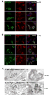
Figure 3. The autophagosome marker GFP-LC3 colocalizes with markers of MHC class II loading compartments in human epithelial cell lines and dendritic cells
(A) MDAMC cells were treated with 200 U/ml IFN-γ, transiently transfected with a GFP-LC3 reporter construct and 48 h later stained with antibodies to MHC class II, HLA-DM, LAMP-2 and DAPI for DNA content. Colocalization of GFP-LC3 with MIIC markers was analyzed by confocal microscopy. Scale bars: 10 μm. Representative cells from one experiment out of three are shown. (B) Same as (A), except that 50 μM chloroquine (CQ) was present during the last 10 h of the culture. Scale bars: 10 μm. Representative cells from one experiment out of three are shown. (C) Colocalization of GFP-LC3 and MHC class II molecules in electron-dense multivesicular compartments. Untreated (I and II) or CQ-treated (III and IV) MDAMC cells stably expressing GFP-LC3 and MHC class II-positive due to IFN-γ induction were fixed in 4% paraformaldehyde and cut into 80 nm-thin cryosections. Sections were labeled with an HLA-DR-specific antiserum and 10 nm protein A-Gold (PAG10) and antibody-PAG complexes were irreversibly fixed with glutaraldehyde. Subsequently, sections were labeled with a GFP-specific antibody and 15 nm protein A-Gold (PAG15) and were analyzed by electron microscopy. As a control, PAG10 and PAG15 were interchanged and were shown to produce the same labeling pattern (I/II vs. III/IV). Insets from panels I and III are shown at higher magnification in panels II and IV, respectively. Representative fields from one experiment out of three are shown.
Figure 4
Figure 4. GFP-LC3 is degraded in MHC class II loading compartments of dendritic cells
(A) GFP-LC3-expressing immature DCs were treated with 50 μM chloroquine for 10 h (+CQ). Cells were stained with an MHC class II-specific antibody and DAPI and colocalization of GFP-LC3 with MHC class II was analyzed by confocal microscopy. Scale bar: 10 μm. Representative cells from one experiment out of three are shown. (B) Same experiment as in (A) was performed with mature DCs. In the majority of mature DCs MHC class II was mainly localized at the cell surface, but a subset of cells had intracellular MHC class II compartments (white arrow). Scale bar: 10 μm. Representative cells from one experiment out of three are shown.
Figure 5
Figure 5. GFP-LC3 minimally colocalizes with markers of early endosomes or MHC class I loading compartments
(A) MDAMC cells were transiently transfected with a GFP-LC3 reporter construct and 36 h later treated with 50 μM chloroquine for 10 h. Cells were stained with antibodies to early endosomal antigen (EEA1) or transferrin receptor (TR) and DAPI and analyzed by confocal microscopy. Scale bars: 10 μm. Representative cells from one experiment out of two are shown. (B) Quantitative analysis for colocalization of GFP-LC3 with MHC class II, HLA-DM and EEA1 in untreated or CQ-treated MDAMC cells. Data represent means from 10–15 cells from one representative experiment out of two. Error bars indicate standard deviations. P-values from homocedastic, one-tailed student’s T test statistics are shown. (C) As in (A), except that cells were stained with an MHC class I-specific antibody.
Figure 6
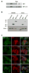
Figure 6. Targeting of influenza A matrix protein 1 (MP1) to autophagosomes by fusion to Atg8/LC3
(A) Schematic diagram of the two constructs encoding for MP1 and MP1-LC3, respectively. The influenza A virus matrix protein 1 (MP1) coding sequence was fused to the N-terminus of the human LC3 sequence, either with or without a stop codon at the 3′ end of MP1. (B) HaCat and MDAMC cell lines were stably transfected with lentiviral MP1 and MP1-LC3 constructs and protein expression was analyzed by Western blot with anti-MP1 antiserum. Actin blot shows equal protein loading. (C) MDAMC cells stably expressing GFP-LC3 were infected with lentivirus encoding for MP1 or MP1-LC3. To inhibit degradation of autophagosome substrates in lysosomes, cells were treated with 50 μM CQ for 10 h (+CQ). Cells were stained with anti-MP1 antiserum and DAPI and analyzed by confocal microscopy. Scale bars: 10 μm. Representative fields from one experiment out of two are shown.
Figure 7
Figure 7. Fusion of MP1 to LC3 enhances CD4+ T cell recognition, while leaving CD8+ T cell recognition unaffected
(A) MP1-specific CD4+ T cell clones 9.26, 10.9 and 11.46 were stimulated at various effector to target cell (ET) ratios with different MP1 or MP1-LC3 expressing target cells: HaCat cells (IFN-γ-treated to induce MHC class II), CM-LCL or immature/mature DCs. The next day, IFN-γ in culture supernatants was measured by ELISA to assess MHC class II presentation of MP1. As a positive control, target cells were pulsed with cognate peptide (+pept.) and as negative controls, untransfected and GFP-LC3-transfected target cells were used. Error bars indicate standard deviations and P-values for paired, one-tailed student’s T test statistics across all E:T ratios are shown. For each target cell type, one representative clone out of three is shown and experiment was performed twice. (B) As in (A), but MP1-specific CD8+ T cell clone 9.2 was used for all experiments. Target cells were MP1 or MP1-LC3 expressing MDAMC cells (IFN-γ-treated or untreated), CM-LCL or immature/mature DCs. Error bars indicate standard deviations. One of two experiments is shown.
Figure 8
Figure 8. Autophagosome targeting depends on covalent coupling of LC3 to the autophagosome membrane via Gly120
(A) MDAMC cells stably expressing GFP-LC3 were infected with lentivirus encoding for MP1, MP1-LC3 or MP1-LC3(G120A). To visualize autophagosomes, cells were treated with 50 μM CQ for 10 h (+CQ) and were stained with anti-MP1 antiserum and DAPI and analyzed by confocal microscopy. Scale bars: 10 μm. (B) The MP1-specific CD4+ T cell clone 11.46 or the MP1-specific CD8+ T cell clone 9.2 were stimulated at various effector to target cell (ET) ratios with target cells expressing either MP1, MP1-LC3 or MP1-LC3(G120). The next day, IFN-γ in culture supernatants was measured by ELISA to assess presentation of MP1 on MHC class II or I, respectively. Error bars indicate standard deviations and P-values for paired, one-tailed student’s T test statistics across all E:T ratios are shown. One of two experiments is shown.

V体育ios版 - Comment in

"V体育ios版" References

    1. Appay V, Zaunders JJ, Papagno L, Sutton J, Jaramillo A, Waters A, Easterbrook P, Grey P, Smith D, McMichael AJ, et al. Characterization of CD4+ CTLs ex vivo. J Immunol. 2002;168:5954–5958. - PubMed
    1. Banchereau J, Steinman RM. Dendritic cells and the control of immunity. Nature. 1998;392:245–252. - PubMed
    1. Berg TO, Fengsrud M, Stromhaug PE, Berg T, Seglen PO. Isolation and characterization of rat liver amphisomes. Evidence for fusion of autophagosomes with both early and late endosomes. J Biol Chem. 1998;273:21883–21892. - PubMed
    1. Bevan MJ. Helping the CD8+ T-cell response. Nat Rev Immunol. 2004;4:595–602. - PubMed
    1. Brazil MI, Weiss S, Stockinger B. Excessive degradation of intracellular protein in macrophages prevents presentation in the context of major histocompatibility complex class II molecules. Eur J Immunol. 1997;27:1506–1514. - PubMed (VSports手机版)

Publication types

MeSH terms

Substances