Olfactory uptake of manganese requires DMT1 and is enhanced by anemia
- PMID: 17116743
- PMCID: "V体育官网" PMC2432183
- DOI: "V体育ios版" 10.1096/fj.06-6710com
Olfactory uptake of manganese requires DMT1 and is enhanced by anemia (V体育平台登录)
Abstract (V体育官网)
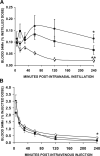
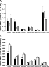
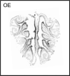
Manganese, an essential nutrient, can also elicit toxicity in the central nervous system (CNS) VSports手机版. The route of exposure strongly influences the potential neurotoxicity of manganese-containing compounds. Recent studies suggest that inhaled manganese can enter the rat brain through the olfactory system, but little is known about the molecular factors involved. Divalent metal transporter-1 (DMT1) is the major transporter responsible for intestinal iron absorption and its expression is regulated by body iron status. To examine the potential role of this transporter in uptake of inhaled manganese, we studied the Belgrade rat, since these animals display significant defects in both iron and manganese metabolism due to a glycine-to-arginine substitution (G185R) in their DMT1 gene product. Absorption of intranasally instilled 54Mn was significantly reduced in Belgrade rats and was enhanced in iron-deficient rats compared to iron-sufficient controls. Immunohistochemical experiments revealed that DMT1 was localized to both the lumen microvilli and end feet of the sustentacular cells of the olfactory epithelium. Importantly, we found that DMT1 protein levels were increased in anemic rats. The apparent function of DMT1 in olfactory manganese absorption suggests that the neurotoxicity of the metal can be modified by iron status due to the iron-responsive regulation of the transporter. .
Figures


References
-
- Barbeau A, Inoue N, Cloutier T. Role of manganese in dystonia. Adv. Neurol. 1976;14:339–352. - PubMed
-
- Barbeau A. Manganese and extrapyramidal disorders (a critical review and tribute to Dr. George C. Cotzias). Neurotoxicology. 1984;5:13–35. - "V体育安卓版" PubMed
-
- Donaldson J. The physiopathologic significance of manganese in brain: its relation to schizophrenia and neurode-generative disorders. Neurotoxicology. 1987;8:451–462. - PubMed
-
- Hinderer RK. Toxicity studies of methylcyclopentadienyl manganese tricarbonyl (MMT). Am. Ind. Hyg. Assoc. J. 1979;40:164–167. - PubMed
-
- Kaiser J. Manganese: a high-octane dispute. Science. 2003;300:926–928. - PubMed (VSports手机版)
Publication types
- Actions (V体育平台登录)
- Actions (VSports手机版)
MeSH terms
- "VSports注册入口" Actions
- Actions (VSports手机版)
- V体育ios版 - Actions
- VSports app下载 - Actions
- "V体育官网入口" Actions
- Actions (V体育2025版)
- Actions (V体育2025版)
- VSports手机版 - Actions
Substances
- Actions (V体育2025版)
- VSports手机版 - Actions
- VSports - Actions
Grants and funding
VSports手机版 - LinkOut - more resources
"VSports注册入口" Full Text Sources
V体育ios版 - Medical

