Skip to main page content
U.S. flag

An official website of the United States government

Dot gov

The . gov means it’s official. Federal government websites often end in . gov or . mil VSports app下载. Before sharing sensitive information, make sure you’re on a federal government site. .

Https

The site is secure V体育官网. The https:// ensures that you are connecting to the official website and that any information you provide is encrypted and transmitted securely. .

. 2006 Nov 27;203(12):2763-77.
doi: 10.1084/jem.20051759. Epub 2006 Nov 20.

"V体育官网入口" An inflammation-induced mechanism for leukocyte transmigration across lymphatic vessel endothelium

Affiliations

"V体育官网" An inflammation-induced mechanism for leukocyte transmigration across lymphatic vessel endothelium

Louise A Johnson (V体育平台登录) et al. J Exp Med. .

Abstract

The exit of antigen-presenting cells and lymphocytes from inflamed skin to afferent lymph is vital for the initiation and maintenance of dermal immune responses. How such an exit is achieved and how cells transmigrate the distinct endothelium of lymphatic vessels are unknown. We show that inflammatory cytokines trigger activation of dermal lymphatic endothelial cells (LECs), leading to expression of the key leukocyte adhesion receptors intercellular adhesion molecule 1 (ICAM-1), vascular cell adhesion molecule 1 (VCAM-1), and E-selectin, as well as a discrete panel of chemokines and other potential regulators of leukocyte transmigration VSports手机版. Furthermore, we show that both ICAM-1 and VCAM-1 are induced in the dermal lymphatic vessels of mice exposed to skin contact hypersensitivity where they mediate lymph node trafficking of dendritic cells (DCs) via afferent lymphatics. Lastly, we show that tumor necrosis factor alpha stimulates both DC adhesion and transmigration of dermal LEC monolayers in vitro and that the process is efficiently inhibited by ICAM-1 and VCAM-1 adhesion-blocking monoclonal antibodies. These results reveal a CAM-mediated mechanism for recruiting leukocytes to the lymph nodes in inflammation and highlight the process of lymphatic transmigration as a potential new target for antiinflammatory therapy. .


Figures
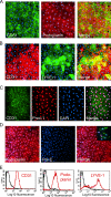
Figure 1.
Figure 1.
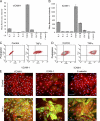
Characteristics of primary HDLECs isolated by LYVE-1 immunoselection. HDLECs isolated from human dermis by LYVE-1 immunomagnetic bead selection are shown after dual immunofluorescence staining for lymphatic and blood vascular-specific markers. (A and B) Confluent monolayers stained for the panendothelial marker CD31 and the lymphatic endothelial markers podoplanin or LYVE-1, respectively. In contrast to podoplanin, which is expressed by all HDLECs, LYVE-1 shows considerable variation, reflecting the heterogeneity seen in normal tissue lymphatics (see Results). (C) Triple staining for CD31, the lymphatic endothelial transcription factor PROX-1, and the nuclear stain DAPI. Note that all cells contain PROX-1–positive nuclei. (D) HDLEC monolayers stained for podoplanin and the blood vascular marker pal-E. Note the complete absence of pal-E–positive cells. Blood vascular endothelial cells positive for pal-E in the mixed endothelial culture preceding LYVE-1 immunoselection are shown for comparison (inset). (E) FACS histograms of HDLECs stained for CD31, podoplanin, or LYVE-1 (red). Isotype-matched controls are shown (black).
Figure 2.
Figure 2.
Inflammatory cytokines up-regulate surface expression of ICAM-1, VCAM-1, and E-selectin in cultured HDLECs. Cells were cultured for 24 h in the presence of individual proinflammatory cytokines or chemokines before immunostaining for either (A) VCAM-1 or (B) ICAM-1 and quantitation by FACS analysis. Data represent the mean ± SEM (n = 3). (C and D) Dot plots show VCAM-1 or ICAM-1 expression in HDLECs cultured in the presence or absence (control) of TNF-α and assessed by dual staining for podoplanin and CAMs. Note that all cells expressing CAMs are positive for podoplanin (top right quadrants). (E) Representative double immunofluorescence micrographs showing induction of CAMs and E-selectin (green) in podoplanin-positive (red) HDLECs, as indicated with nuclei counterstained for DAPI. Bar, 50 μm.
Figure 3.
Figure 3.
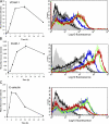
Kinetics of TNF-induced CAM and E-selectin expression in cultured primary HDLECs. (A–C) Respective time courses for induction of VCAM-1, ICAM-1, and E-selectin in HDLECs cultured for 0–48 h in the presence or absence of 1 ng/ml TNF-α, as assessed by FACS analysis. Representative histograms are shown for cells stained with isotype-matched control Ig (light gray) or mAbs to the appropriate adhesion molecules in unstimulated cells (black) or cells treated with TNF-α for 3 h (dark gray), 6 h (blue), 12 h (green), and 24 h (red).
Figure 4.
Figure 4.
TNF-α induces proinflammatory chemokine production in cultured primary HDLECs. Cells were stimulated with 1 ng/ml TNF-α over a time course of 48 h, and concentrations of MCP-1, RANTES, and MIP-3α secreted into the culture supernatant were measured by ELISA. Data represent the mean ± SEM (n = 3).
Figure 5.
Figure 5.
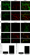
In vivo expression of ICAM-1 and VCAM-1 in mouse dermal lymphatics induced by skin contact hypersensitivity. Skin inflammation was induced in mouse ear by sensitization and subsequent challenge with oxazolone before analysis of lymphatic vessel CAM expression by immunofluorescence microscopy. (A and B) Whole-mount sections of oxazolone-challenged and contralateral-unchallenged (control) ears dual-stained for podoplanin (green) and ICAM-1 or VCAM-1, respectively (red). Note the weak expression of ICAM-1 confined to podoplanin-negative (blood) vessels in uninflamed skin (A) and the focal up-regulation of both ICAM-1 and VCAM-1 on podoplanin-positive (lymphatic) vessels in inflamed skin (A and B). Images were captured by confocal microscopy. Bars, 100 μm. (C and D) Quantitative estimates for the numbers of ICAM-1+/podoplanin+ and VCAM-1+/podoplanin+ vessels determined by counting 21 separate fields of view (7 fields/mouse) in control and oxazolone-treated ear sections. Data represent the mean ± SEM.
Figure 6.
Figure 6.
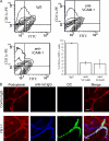
In vivo trafficking of skin DCs via afferent lymphatics is dependent on ICAM-1 and VCAM-1 adhesion. The involvement of ICAM-1 and VCAM-1 in the trafficking of DCs via afferent lymphatics was investigated in mice with oxazolone-induced skin hypersensitivity. (A) Recoveries of FITC+/CD11c+ skin DCs in the draining lymph nodes 24 h after FITC skin painting of oxazolone-sensitized mice that received prior injection of neutralizing mAbs to VCAM-1, ICAM-1, or control rat Ig. Data represent the mean recoveries ± SEM (obtained from three separate experiments). (B) To show retention of DCs within the skin, CMFDA-labeled bone marrow–derived DCs from a littermate were intradermally injected into the ear tissue of sensitized mice that received prior injection of a neutralizing mAb to ICAM-1 (YN1-1) or control rat Ig. After 24 h, ears were removed, and whole-mount staining was performed using antipodoplanin with Alexa Fluor 568 (red) and Cy5-conjugated goat anti–rat Cy5 (blue) to detect binding of neutralizing antibody within the tissue. Bars, 100 μm.
Figure 7.
Figure 7.
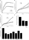
MDDC transmigration of TNF-α–stimulated HDLEC monolayers is dependent on ICAM-1 and VCAM-1. Transmigration of Cell Tracker Green fluorescently labeled MDDCs across either unstimulated or TNF-α–stimulated HDLEC monolayers plated on the undersurface of Fluoroblok filters was monitored in the presence or absence of selected adhesion blocking antibodies over a 12-h period. Progress curves are shown for MDDC transmigration across (A) control unstimulated versus TNF-α–stimulated HDLECs, (B) TNF-α–stimulated HDLECs treated with control rat IgG versus VCAM-1–neutralizing mAb P8B1, and (C) TNF-α–stimulated HDLECs treated with control rat IgG versus ICAM-1– neutralizing mAb P2A4. Data represent the mean ± SEM (n = 4). (D) Comparative effects of individual ICAM-1 mAbs 15.2 and P2A4, VCAM-1 mAbs 51-10C9 and P8B1, ICAM-1 mAb 15.2 and VCAM-1 mAb P8B1 together, the LFA-1 mAb 24, and control mouse IgG on MDDC transmigration of TNF-α–stimulated HDLECs. The level of transmigration across unstimulated HDLECs is shown for comparison. Data from three independent experiments are normalized to the measured levels of transmigration in the presence of control IgG (100% maximal transmigration) in each case and represent the mean ± SEM (n = 4). (E) Permeability of confluent HDLEC monolayers to unconjugated Alexa Fluor 488 measured as dye recovered in the lower chamber of Fluoroblok filter wells after a 6-h incubation at 37°C. Data represent the mean ± SEM.
Figure 8.
Figure 8.
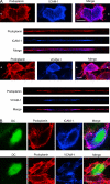
VCAM-1 and ICAM-1 are expressed on both luminal and basolateral surfaces of HDLECs. Cells were cultured on clear-membrane inserts and stimulated with TNF-α for 24 h before staining with antipodoplanin (red) and either anti–ICAM-1 or anti–VCAM-1 (blue) and analysis by confocal microscopy. Staining was performed either on HDLECs cultured (A) in the absence of MDDCs or (B) at 10 h after addition of Cell Tracker Green fluorescently labeled MDDCs. (A, top) Asterisks depict the axis through which individual LECs were imaged in z-section (bottom). Bars, 10 μm.
Figure 9.
Figure 9.
MDDC adhesion to TNF-α–stimulated HDLECs is dependent on ICAM-1 and VCAM-1. Cell Tracker Green fluorescently labeled MDDCs were applied to TNF-α–stimulated HDLEC monolayers plated in 24-well plates, preincubated with either IgG control or ICAM-1 mAbs (15.2 or P2A4) or VCAM-1 mAbs (P8B1 or IGII), in triplicate. At 3 and 10 h, the numbers of adherent MDDCs were measured. Representative data from three independent experiments are shown and represent the mean ± SEM (n = 3).

References

    1. Mellman, I., and R.M. Steinman. 2001. Dendritic cells: specialized and regulated antigen processing machines. Cell. 106:255–258. - V体育官网 - PubMed
    1. Muller, W.A. 2003. Leukocyte-endothelial cell interactions in leukocyte transmigration and the inflammatory response. Trends Immunol. 24:327–334. - PubMed
    1. Randolph, G.J., V. Angeli, and M.A. Swartz. 2005. Dendritic cell trafficking to lymph nodes though lymphatic vessels. Nat. Rev. Immunol. 5:617–628. - "V体育平台登录" PubMed
    1. Sallusto, F., P. Schaerli, P. Loetscher, C. Schaniel, D. Lenig, C.R. Mackay, S. Qin, and A. Lanzavecchia. 1998. Rapid and co-ordinated switch in chemokine receptor expression during dendritic cell maturation. Eur. J. Immunol. 28:2760–2769. - PubMed
    1. Saeki, H., A.M. Moore, M.J. Brown, and S.T. Hwang. 1999. Secondary lymphoid tissue chemokine (SLC) and CC chemokine receptor 7 (CCR7) participate in the emigration pathway of mature dendritic cells from the skin to regional lymph nodes. J. Immunol. 162:2472–2475. - PubMed

Publication types

MeSH terms

Substances (V体育2025版)