Skip to main page content
U.S. flag

An official website of the United States government

Dot gov

The . gov means it’s official. Federal government websites often end in . gov or VSports app下载. mil. Before sharing sensitive information, make sure you’re on a federal government site. .

Https

The site is secure. The https:// ensures that you are connecting to the official website and that any information you provide is encrypted and transmitted securely V体育官网. .

. 2006 Nov 15;20(22):3147-60.
doi: 10.1101/gad.1475506.

Aggressive pancreatic ductal adenocarcinoma in mice caused by pancreas-specific blockade of transforming growth factor-beta signaling in cooperation with active Kras expression

Affiliations

Aggressive pancreatic ductal adenocarcinoma in mice caused by pancreas-specific blockade of transforming growth factor-beta signaling in cooperation with active Kras expression

Hideaki Ijichi et al. Genes Dev. .

V体育官网 - Abstract

Pancreatic ductal adenocarcinoma (PDAC) is an almost uniformly lethal disease in humans. Transforming growth factor-beta (TGF-beta) signaling plays an important role in PDAC progression, as indicated by the fact that Smad4, which encodes a central signal mediator downstream from TGF-beta, is deleted or mutated in 55% and the type II TGF-beta receptor (Tgfbr2) gene is altered in a smaller subset of human PDAC. Pancreas-specific Tgfbr2 knockout mice have been generated, alone or in the context of active Kras (Kras(G12D)) expression, using the Cre-loxP system driven by the endogenous Ptf1a (pancreatic transcription factor-1a) locus VSports手机版. Pancreas-selective Tgfbr2 knockout alone gave no discernable phenotype in 1. 5 yr. Pancreas-specific Kras(G12D) activation alone essentially generated only intraepithelial neoplasia within 1 yr. In contrast, the Tgfbr2 knockout combined with Kras(G12D) expression developed well-differentiated PDAC with 100% penetrance and a median survival of 59 d. Heterozygous deletion of Tgfbr2 with Kras(G12D) expression also developed PDAC, which indicated a haploinsufficiency of TGF-beta signaling in this genetic context. The clinical and histopathological manifestations of the combined Kras(G12D) expression and Tgfbr2 knockout mice recapitulated human PDAC. The data show that blockade of TGF-beta signaling and activated Ras signaling cooperate to promote PDAC progression. .


Figures
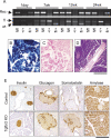
Figure 1.
Figure 1.
Normal pancreas development in Tgfbr2 knockout mice. (A) PCR detected Tgfbr2 allele recombination in the pancreas tissues of 1-d to 24-wk-old mice. (Top) PCR to detect Tgfbr2 allele recombination. The recombined allele was detected in the Ptf1acre/+;Tgfbr2flox/flox and Ptf1acre/+;Tgfbr2flox/f+ mice throughout the ages examined. (Bottom) Genotyping PCR to detect floxed and wild-type Tgfbr2 alleles. Some floxed alleles appeared to be diminished in those mice that showed the allele recombination. (Rec) Recombined allele; (fl) floxed allele; (wt) wild-type allele of Tgfbr2 gene; (fl/fl) Ptf1acre/+;Tgfbr2flox/flox mice; (fl/+) Ptf1acre/+;Tgfbr2flox/f+ mice; (+/+) Tgfbr2flox/flox mice. (B,C) β-Galactosidase in situ staining. Cre recombination was homogeneously observed all through the pancreas of Ptf1acre/+;Tgfbr2flox/flox;Rosa26r mice (B), but not in that of Tgfbr2flox/flox;Rosa26r mice (C). (D) H&E staining of 1.5-yr-old pancreas of the Ptf1acre/+;Tgfbr2flox/flox mouse. The pancreas was normally developed and has no abnormal findings in Tgfbr2 knockout mice. (E) Immunohistochemistry of endocrine and exocrine pancreas markers at 7 wk of age. Insulin, glucagon, and amylase staining showed no difference. Somatostatin expression was more prominent in the Tgfbr2 knockout mice at this stage. (Control) Tgfbr2flox/flox mice; (Tgfbr2 KO) Ptf1acre/+;Tgfbr2flox/flox mice. Bars: B,C,E 250 μm; D, 500 μm.
Figure 2.
Figure 2.
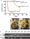
KrasG12D expression plus Tgfbr2 knockout developed aggressive pancreatic tumors. (A) Kaplan-Meyer curve. Ptf1acre/+;LSL-KrasG12D/+;Tgfbr2flox/flox mice (Kras + Tgfbr2KO) showed a dramatically shortened survival (median survival: 59 d). Ptf1acre/+;LSL-KrasG12D/+; Tgfbr2flox/+ mice (Kras + Tgfbr2L/+) also showed a shortened survival compared with the controls after 200 d. The control group includes the genotypes of LSL-KrasG12D/+;Tgfbr2flox/flox, Ptf1acre/+;Tgfbr2flox/flox, Ptf1acre/+, and Ptf1acre/+;LSL-KrasG12D/+. (B,C) Gross images of the pancreatic tumor from Ptf1acre/+;LSL-KrasG12D/+;Tgfbr2flox/flox mice. The abdomen was distended due to the tumor (arrowhead) and ascites. (B) Note the bloody ascites in this mouse. The tumor (arrowhead) occupied the entire pancreas and compressed adjacent organs. (C) Note the apparent jaundice in this mouse. (D) PCR detected recombination of the LSL-KrasG12D and floxed Tgfbr2 alleles in a pancreas-specific manner. (Rec) Recombined allele; (wt) wild-type allele. The genotypes of mice are K133, K59, K131, K143: Ptf1acre/+;LSL-KrasG12D/+;Tgfbr2flox/flox; K127: Ptf1acre/+;LSL-KrasG12D/+; K130: Ptf1acre/+; Tgfbr2flox/flox; K129: LSL-KrasG12D/+; Tgfbr2flox/flox; K125: wild-type. (P) Pancreas; (L) liver.
Figure 3.
Figure 3.
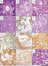
Histology of the Ptf1acre/+;LSL-KrasG12D/+;Tgfbr2flox/flox pancreatic tumors. Ptf1acre/+;LSL-KrasG12D/+ (A,H,J) and Ptf1acre/+;LSL-KrasG12D/+;Tgfbr2flox/flox (BG,I,K,L) pancreas (7–9 wk of age). (A–D) H&E staining. (A) The Ptf1acre/+;LSL-KrasG12D/+ mice showed mPanIN lesions surrounded by stromal capsule and still abundant normal pancreas areas. (B) The Ptf1acre/+;LSL-KrasG12D/+;Tgfbr2flox/flox pancreas showed well-differentiated ductal adenocarcinoma, with a rich stromal component. Some areas still retained nuclear polarity and abundant cytoplasm (C); however, others showed nuclear atypia and a high nuclear–cytoplasmic ratio (D). (E) Pan-cytokeratin staining revealed tumor epithelial cells invading into the stromal regions. Positive alcian blue (F) and CK-19 staining (G) confirmed the mucin-producing (blue), ductal phenotype of the tumor. Vimentin (H,I) and smooth muscle actin (J,K) staining was negative in the tumor epithelia, whereas prominent stromal infiltration (strong positive) was observed. (L) H&E staining of the Ptf1acre/+; LSL-KrasG12D/+;Tgfbr2flox/flox tumor also showed acinar–ductal metaplasia lesions. The arrows indicate acinar–ductal metaplasia (ductal lesions containing acinar granules). The arrowheads indicate an mPanIN-1B lesion. Bars: B,E, 250 μm; A, C,D,FL, 125 μm.
Figure 4.
Figure 4.
Immunostaining of the Ptf1acre/+;LSL-KrasG12D/+;Tgfbr2flox/flox and Ptf1acre/+;LSL-KrasG12D/+ pancreatic tumors. EGFR and phospho-ERK were highly expressed both in the Ptf1acre/+;LSL-KrasG12D/+ mPanIN lesions (top panels) and Ptf1acre/+;LSL-KrasG12D/+; Tgfbr2flox/flox PDAC lesions (bottom panels). erbB2/Her2 was also expressed in both lesions; however, more advanced stage lesions of the latter showed weaker or negative staining. E-cadherin immunofluorescence revealed a regular cell-junctional staining in the mPanIN lesions, whereas the PDAC lesions frequently showed nonjunctional, disorganized E-cadherin expression. (Green) E-Cadherin; (blue) DAPI. (Kras PanIN) mPanIN in the Ptf1acre/+;LSL-KrasG12D/+/+ mice; (Kras + RIIKO PDAC) PDAC in the Ptf1acre/+; LSL-KrasG12D/+;Tgfbr2flox/flox mice. Bars, 125 μm.
Figure 5.
Figure 5.
Molecular analysis of the Ptf1acre/+;LSL-KrasG12D/+; Tgfbr2flox/flox PDAC cell lines. (A) Western blot analysis using total cell lysates of the established pancreatic cell lines from the Ptf1acre/+;LSL-KrasG12D/+;Tgfbr2flox/flox PDAC (Kras + Tgfbr2KO cells) and Ptf1acre/+;LSL-KrasG12D/+ pancreas (Kras cells) revealed intact Smad4, p53 protein expression in most of the cell lines and loss of TβRII only in the Kras + Tgfbr2KO cell lines. p16INK4a protein was variably expressed even in the Kras cell lines. (Actin) Loading control. (B) Analysis of p53 function by γ-irradiation. p53 protein was induced by γ-irradiation in all cell lines examined. (Tubulin) Loading control. (C) RT–PCR for p16INK4a and p19ARF mRNA expression. p16INK4a mRNA level was variable in the cell lines, which was well correlated with the protein level. The p19ARF mRNA level was relatively consistent with that of p16INK4a. (GAPDH) Loading control. (D) TGF-β–Smad signaling analysis. TGF-β-induced Smad3 phosphorylation was found only in the Kras cells (TβRII-intact), but was completely lost in all the Kras + Tgfbr2KO cells. (Actin) Loading control.
Figure 6.
Figure 6.
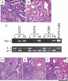
Haploinsufficiency of Tgfbr2 in the context of KrasG12D expression promoted PDAC progression. (A,B) H&E staining of 32-wk-old Ptf1acre/+; LSL-KrasG12D/+;Tgfbr2flox/+ mouse pancreas. (A) Well-differentiated ductal adenocarcinoma in the pancreas head region. (B) mPanIN lesions were dominant in the pancreas tail region. (C) LOH analysis by PCR after laser-capture microdissection. DNA was extracted from the primary pancreas tumor epithelia, metastatic liver tumor epithelia, normal liver, or normal duodenum of the Ptf1acre/+;LSL-KrasG12D/+;Tgfbr2flox/+ mice, using laser-capture microdissection. Subsequent PCR showed that all primary pancreas tumor and one metastatic liver tumor epithelia clearly retained the wild-type Tgfbr2 allele (K372 P, K374 P and M, K388 P1 and P2), indicating a lack of LOH. (K372, K374, K388) Ptf1acre/+; LSL-KrasG12D/+;Tgfbr2flox/+ mice; (K375 cells) a cell line of Ptf1acre/+;LSL-KrasG12D/+;Tgfbr2flox/flox mouse PDAC. (L) Normal liver; (D) normal duodenum; (P) primary pancreas tumor epithelia; (M) metastatic liver tumor epithelia; (P1) well-differentiated PDAC epithelia; (P2) poorly differentiated PDAC epithelia; (Rec) recombined; (fl) floxed; (wt) wild-type Tgfbr2 allele. (D–F) H&E staining of 50-wk-old Ptf1acre/+;LSL-KrasG12D/+;Tgfbr2flox/+ mouse pancreas and liver. The tumor showed sarcomatoid histology (s) along with ductal adenocarcinoma lesions (arrow) (D), and both sarcomatoid (s) and ductal tumor (arrow) directly invaded into the liver parenchyma (L) (E,F). Bars: E, 250 μm; AD,F, 125 μm.
Figure 7.
Figure 7.
CTGF overexpression in the stroma of Ptf1acre/+;LSL-KrasG12D/+;Tgfbr2flox/flox PDAC. CTGF immunohistochemistry of 7-wk-old Ptf1acre/+;LSL-KrasG12D/+ (A,B) and Ptf1acre/+;LSLKrasG12D/+;Tgfbr2flox/flox (C,D) mouse pancreas. (A) The Ptf1acre/+; LSL-KrasG12D/+ pancreas stroma shows negative staining of CTGF. (C,D) The Ptf1acre/+;LSL-KrasG12D/+;Tgfbr2flox/flox PDAC stroma demonstrated strong expression of CTGF. Note that the stroma adjacent to the tumor epithelia especially exhibits high CTGF expression. Bars: C, 500 μm; A, 250 μm; B,D, 125 μm.

VSports最新版本 - References

    1. Aguirre, A.J., Bardeesy, N., Sinha, M., Lopez, L., Tuveson, D.A., Horner, J., Redston, M.S., DePinho, R.A. Activated Kras and Ink4a/Arf deficiency cooperate to produce metastatic pancreatic ductal adenocarcinoma. Genes & Dev. 2003;17:3112–3126. - PMC - PubMed
    1. Aikawa, T., Gunn, J., Spong, S.M., Klaus, S.J., Korc, M. Connective tissue growth factor-specific antibody attenuates tumor growth, metastasis, and angiogenesis in an orthotopic mouse model of pancreatic cancer. Mol. Cancer Ther. 2006;5:1108–1116. - PubMed
    1. Bardeesy, N., Aguirre, A.J., Chu, G.C., Cheng, K.H., Lopez, L.V., Hezel, A.F., Feng, B., Brennan, C., Weissleder, R., Mahmood, U., et al. Both p16(Ink4a) and the p19(Arf)–p53 pathway constrain progression of pancreatic adenocarcinoma in the mouse. Proc. Natl. Acad. Sci. 2006;103:5947–5952. - PMC - PubMed
    1. Bierie, B., Moses, H.L. TGF-β and cancer. Cytokine Growth Factor Rev. 2006;17:29–40. - PubMed
    1. Bottinger, E.P., Jakubczak, J.L., Roberts, I.S., Mumy, M., Hemmati, P., Bagnall, K., Merlino, G., Wakefield, L.M. Expression of a dominant-negative mutant TGF-β type II receptor in transgenic mice reveals essential roles for TGF-β in regulation of growth and differentiation in the exocrine pancreas. EMBO J. 1997;16:2621–2633. - PMC - PubMed

V体育官网入口 - Publication types

MeSH terms

Substances