Skip to main page content
U.S. flag

An official website of the United States government

Dot gov

The . gov means it’s official. Federal government websites often end in VSports app下载. gov or . mil. Before sharing sensitive information, make sure you’re on a federal government site. .

Https

The site is secure V体育官网. The https:// ensures that you are connecting to the official website and that any information you provide is encrypted and transmitted securely. .

. 2006 Nov 28;103(48):18238-42.
doi: 10.1073/pnas.0607057103. Epub 2006 Nov 15.

"V体育官网入口" Human cancers express a mutator phenotype

Affiliations

"VSports app下载" Human cancers express a mutator phenotype

"VSports app下载" Jason H Bielas et al. Proc Natl Acad Sci U S A. .

Erratum in

  • Proc Natl Acad Sci U S A. 2006 Dec 19;103(51):19605

Abstract

Cancer cells contain numerous clonal mutations, i. e. , mutations that are present in most or all malignant cells of a tumor and have presumably been selected because they confer a proliferative advantage. An important question is whether cancer cells also contain a large number of random mutations, i. e. , randomly distributed unselected mutations that occur in only one or a few cells of a tumor. Such random mutations could contribute to the morphologic and functional heterogeneity of cancers and include mutations that confer resistance to therapy. We have postulated that malignant cells exhibit a mutator phenotype resulting in the generation of random mutations throughout the genome. We have recently developed an assay to quantify random mutations in human tissue with unprecedented sensitivity. Here, we report measurements of random single-nucleotide substitutions in normal and neoplastic human tissues. In normal tissues, the frequency of spontaneous random mutations is exceedingly low, less than 1 x 10(-8) per base pair VSports手机版. In contrast, tumors from the same individuals exhibited an average frequency of 210 x 10(-8) per base pair, an elevation of at least two orders of magnitude. Our data document tumor heterogeneity at the single-nucleotide level, indicate that accelerated mutagenesis prevails late into tumor progression, and suggest that elevation of random mutation frequency in tumors might serve as a novel prognostic indicator. .


"VSports app下载" Conflict of interest statement

The authors declare no conflict of interest.

"V体育官网" Figures

Fig. 1.
Fig. 1.
The RMC applied to human tissue. Genomic DNA is isolated from intact tissue and digested with restriction enzymes that do not cut the mutational target sequence, a 4-bp TaqI restriction site (TCGA) in intron VI of p53; blue lines represent the wild-type target sequence, and red lines represent mutant target sequence. A complementary probe (gray lines) that contains dUMP in place of dTMP and a biotinylated nucleotide at the 5′ terminus is hybridized to the mutational target. The hybridized target is isolated by complexing to magnetic beads, digested with TaqI (cleaving the TCGA target site in the wild-type sequence and failing to cleave if a nucleotide substitution is present at that site), and denatured. Rehybridization and TaqI digestion are carried out four times. The probe is then disabled for further hybridization by digestion with uracil-DNA glycosylase, and the mutational target is diluted in 96-well plates so that 1 in ≈10 wells contains a PCR-amplifiable product (red wells) as measured with SYBR green by using real-time QPCR. The mutation frequency is quantified by QPCR amplification and is calculated as the number of wells containing a mutant sequence divided by the product of the total number of target molecules screened and the restriction site length (bp). The mutant sequence of the amplified product in all positive wells is verified by DNA sequencing; C4 and G8 represent the mutant sequences found in wells C4 and G8, respectively, of the 96-well plate shown. In some cases, preliminary verification was carried out by redigestion with TaqI.
Fig. 2.
Fig. 2.
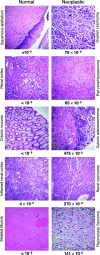
Matched normal and neoplastic tissues analyzed in the RMC assay. Hematoxylin/eosin-stained sections of the paired normal (Left) and tumor (Right) tissues listed in Table 1 are shown. The tissues are normal squamous vaginal epithelium and high-grade papillary serous ovarian carcinoma with psammoma bodies; normal renal cortex and dedifferentiated sclerosing perirenal liposarcoma; normal colonic mucosa and invasive colonic adenocarcinoma; renal cortex with lymphocytic inflammation and malignant renal epithelioid angiomyolipoma; and normal skeletal muscle and high-grade malignant fibrous histiocytoma pleomorphic sarcoma. Immunohistochemical analysis of MLH1 and MSH2 proteins involved in DNA mismatch repair showed the colonic adenocarcinoma to lack MLH1 expression and to have normal expression of MSH2. These results suggest that the tumor is defective in mismatch repair. The mutation frequencies measured in the RMC assay are indicated below each section.
Fig. 3.
Fig. 3.
Tumor mutation spectrum. DNA sequencing of all mutants recovered from tumors showed that C·G to T·A transitions were the most common mutation, and the sequencing permitted distinction between independent random mutational events (gray bars, mutation observed only once) and expansion of mutant clones (white bars, same mutation recovered more than once in the same tumor). Identical mutations observed more than once (expanded mutations) are recorded as one single event; the number of these minority events is indicated by the relatively short length of the white bar extending past the gray bar.

Comment in

References

    1. Weinberg RA. Biology of Cancer. New York: Garland Science; 2007.
    1. Levine AJ. Cell. 1997;88:323–331. - PubMed
    1. Hussain SP, Amstad P, Raja K, Ambs S, Nagashima M, Bennett WP, Shields PG, Ham A-J, Swenberg JA, Marrogi AJ, Harris CC. Cancer Res. 2000;60:3333–3337. - PubMed
    1. Barbacid M. Annu Rev Biochem. 1987;56:779–827. - PubMed
    1. Albertson DG, Collins C, McCormick F, Gray JW. Nat Genet. 2003;34:369–376. - PubMed

Publication types (V体育2025版)

"V体育ios版" LinkOut - more resources