Skip to main page content
U.S. flag

An official website of the United States government

Dot gov

The . gov means it’s official. Federal government websites often end in . gov or . mil. Before sharing sensitive information, make sure you’re on a federal government site. VSports app下载.

Https

The site is secure V体育官网. The https:// ensures that you are connecting to the official website and that any information you provide is encrypted and transmitted securely. .

. 2007 Jan;27(1):324-39.
doi: 10.1128/MCB.01394-06. Epub 2006 Oct 30.

Dual role of SnoN in mammalian tumorigenesis

Affiliations

"V体育安卓版" Dual role of SnoN in mammalian tumorigenesis

Qingwei Zhu et al. Mol Cell Biol. 2007 Jan.

Abstract (VSports手机版)

SnoN is an important negative regulator of transforming growth factor beta signaling through its ability to interact with and repress the activity of Smad proteins. It was originally identified as an oncoprotein based on its ability to induce anchorage-independent growth in chicken embryo fibroblasts. However, the roles of SnoN in mammalian epithelial carcinogenesis have not been well defined. Here we show for the first time that SnoN plays an important but complex role in human cancer. SnoN expression is highly elevated in many human cancer cell lines, and this high level of SnoN promotes mitogenic transformation of breast and lung cancer cell lines in vitro and tumor growth in vivo, consistent with its proposed pro-oncogenic role VSports手机版. However, this high level of SnoN expression also inhibits epithelial-to-mesenchymal transdifferentiation. Breast and lung cancer cells expressing the shRNA for SnoN exhibited an increase in cell motility, actin stress fiber formation, metalloprotease activity, and extracellular matrix production as well as a reduction in adherens junction proteins. Supporting this observation, in an in vivo breast cancer metastasis model, reducing SnoN expression was found to moderately enhance metastasis of human breast cancer cells to bone and lung. Thus, SnoN plays both pro-tumorigenic and antitumorigenic roles at different stages of mammalian malignant progression. The growth-promoting activity of SnoN appears to require its ability to bind to and repress the Smad proteins, while the antitumorigenic activity can be mediated by both Smad-dependent and Smad-independent pathways and requires the activity of small GTPase RhoA. Our study has established the importance of SnoN in mammalian epithelial carcinogenesis and revealed a novel aspect of SnoN function in malignant progression. .


Figures

FIG. 1.
FIG. 1.
Expression of SnoN is elevated in human cancer cell lines. (A) Expression levels of SnoN and Ski are regulated differently in human cancer cell lines. Endogenous SnoN or Ski was isolated by immunoprecipitation from equal amounts of cell lysates prepared from the following cell lines: human breast cancer cell lines (MDA-MB-231, MDA-MB-435, and ZR75b), lung adenocarcinoma lines (A427, A549, and SW1271), melanoma (A375), osteosarcoma (HT1080), and two untransformed human mammary epithelial cell lines (HMT3522-S1 and MCF10A). Immune complexes were analyzed by Western blotting with anti-SnoN or anti-Ski. (B) The snoN mRNA is upregulated in some cancer cell lines. Twenty micrograms of total RNA was isolated from cancer cell lines described in panel A, and snoN expression was analyzed by Northern blotting. 28S and 18S RNA are shown as loading controls. (C) TGF-β-induced phosphorylation of Smad2 and Smad3 in cancer cell lines. Cells were serum starved for 16 h and treated with 100 pM TGF-β1 for the indicated period of time. Equal amounts of total cell lysates were analyzed by Western blotting using antibodies against phospho-Smad2, phospho-Smad3 (P-Smad2 and P-Smad3; top two panels), Smad2 and Smad3 (middle panels), or α-tubulin as a loading control (bottom panel).
FIG. 2.
FIG. 2.
Reduction of SnoN expression restores TGF-β responses in A549 and MDA-MB-231 cancer cell lines. (A) Reduction of snoN expression in A549 lung cancer cells and MDA-MB-231 breast cancer cells by shRNA. Stable shSnoN cell lines expressing the shRNA for human snoN were generated as described in Materials and Methods. SnoN expression in the representative stable clones was assessed by immunoprecipitation and Western blotting and compared with that in parental cells. The levels of α-tubulin in cell lysates were measured by Western blotting and included as a loading control. (B) Phosphorylation of R-Smads in response to TGF-β in A549 and MDA-MB-231 parental and shSnoN cell lines. Cells were treated with 100 pM TGF-β1 for the indicated time periods. Equal amounts of total cell lysates were analyzed by Western blotting using antibodies against phospho-Smad2 (P-Smad2; top panel), phospho-Smad3 (P-Smad3; middle panel) or total Smad2 and Smad3 (bottom panel). (C) Effects of TGF-β on the growth of parental cancer cells and their derived shSnoN cells. Parental A549 cells, MDA-MB-231 cells, or their shRNA-expressing derivatives were treated with increasing concentrations of TGF-β1 and cultured for 4 days. The growth of cells was determined by cell counting, and the number is expressed as a percentage of the number of cells in unstimulated samples. (D) SnoN-deficient cells exhibit increased expression of TGF-β-responsive genes. Parental A549 cells, MDA-MB-231 cells, and their shSnoN derivatives were serum starved for 16 h and stimulated with 100 pM TGF-β1 for 3 h. Total RNA was isolated from these cells, and expression of PAI-1 and p21 was assessed by Northern blotting. 28S and 18S RNA were included as loading controls.
FIG. 3.
FIG. 3.
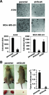
Reducing SnoN expression suppresses the transformed phenotype of cancer cells. (A) Reducing SnoN expression reverses transformation of A549 and MDA-MB-231 cancer cells. Parental A549 or MDA-MB-231 cells and their derived shSnoN cells were subjected to a soft agar colony assay as described in Materials and Methods. The soft agar plates were stained with 0.5 mg/ml MTT and scanned. A representative 1-cm2 area of each six-well plate is shown in the top panel. The number of soft agar colonies in each plate was quantified and is summarized in the bottom panel. (B) Reducing SnoN expression blocks tumorigenicity in vivo. Parental or shSnoN-expressing A549 cells were subcutaneously injected into both flanks of five nude mice. A representative nude mouse at 8 weeks after injection with either parental cells or shSnoN cells is shown in the left panel (top). The tumor size of shSnoN-expressing A549 cells was significantly reduced compared with that of the parental A549 tumor (left panel, bottom). The mean tumor volume was calculated as described in Materials and Methods and graphed in the right panel. Each bar represents the mean ± SEM from 10 primary tumors.
FIG. 4.
FIG. 4.
Reducing SnoN expression enhances EMT. (A) Reducing SnoN expression increases cell motility. Confluent cell monolayers of parental or shSnoN-expressing A549 and MDA-MB-231 cells were wounded with a pipette tip. Wound closure was monitored by microscopy at the indicated times (left panel). Percent wound closure (plotted in right panel) was calculated by measuring the wound closure distance, and this value is expressed as a percentage of the initial wound length. (B) SnoN-deficient A549 cells exhibit increased expression of fibronectin (FN). Parental and shSnoN-expressing A549 cells were treated with 100 pM TGF-β1 for 24 h. Equal amounts of cell lysates prepared from TGF-β-treated and untreated cells were analyzed by Western blotting with antifibronectin. α-Tubulin expression was used as a loading control. (C) Cells expressing shSnoN show increased matrix metalloprotease activity. Parental A549 cells, MDA-MB-231 cells, and their shSnoN-expressing derivatives were cultured on coverslips coated with gelatin-Alexa 488 (to assay MMP2 and MMP9 activity) in the absence or presence of 100 pM TGF-β1 alone or in combination with 10 μM MMP inhibitor GM6001 for 24 to 48 h before processing for immunofluorescence microscopy. Representative in situ zymography micrographs are shown on the left panel. The levels of protease activity within a specified field were quantified by calculating the percentage of digested area normalized to the number of cells within this field and are expressed as the percent digested area per cell (right panel). (D) Reduced expression of SnoN increases stress fiber formation in A549 and MDA-MB-231 cancer cells. Parental A549 cells, MDA-MB-231 cells, and their shSnoN-expressing derivatives were cultured in the absence or presence of 100 pM TGF-β1 for 2 days and then processed for F-actin staining using rhodamine-phalloidin. (E) E-cadherin expression is diminished in SnoN-deficient cells. Parental and shSnoN-expressing A549 cells were treated with 100 pM TGF-β1 for 24 h. Equal amounts of cell lysates prepared from TGF-β-treated and untreated cells were analyzed by Western blotting with anti-E-cadherin. α-Tubulin expression was used as a loading control.
FIG. 5.
FIG. 5.
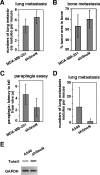
Effects of downregulation of SnoN on tumor metastasis in vivo. (A) Effects of abrogation of SnoN expression on the lung metastasis of MDA-MB-231 cells. Parental or shSnoN-expressing MDA-MB-231 cells were injected into the left cardiac ventricle of 4-week-old anesthetized female nude mice. Lung metastasis assay was carried out as described in Materials and Methods. The results are expressed as mean ± SEM from eight parental cell-injected mice and seven shSnoN cell-injected mice. (B) Effect of abrogation of SnoN expression on the bone metastasis of MDA-MB-231 cells. The bone metastatic potential is indicated by the histomorphometric measurement of tumor area/burden (in percentage) in the cancellous regions of the right distal femora beginning 100 μm below the growth plates. The area of measurement was 1.5 mm in length, and the width was determined by the inside edges of the cortical bone. The results are expressed as means ± SEM from eight parental cell-injected mice and seven shSnoN cell-injected mice. (C) Paraplegia assay in the parental and shSnoN-expressing MDA-MB-231 cell-injected mice. To detect paraplegia, a wire hang test was performed as described in Materials and Methods. The results are expressed as means ± SEM from six parental cell-injected mice and seven shSnoN cell-injected mice. (D) A549 lung cancer metastasis assay. Parental or shSnoN-expressing A549 cells were injected into the tail veins of 4-week-old female nude mice. After 3 months, 10 mice from each group were euthanized and analyzed as described in Materials and Methods. The number of surface tumor nodules in lungs of each mouse was counted. (E) Twist1 expression is upregulated in shSnoN-expressing cells. RNA was extracted from parental or shSnoN-expressing A549 cells. An RT-PCR assay was carried out to detect the expression levels of Twist1 in both parental and shSnoN-expressing A549 cells. RT-PCR with primers amplifying the GAPDH locus is shown as a loading control.
FIG. 6.
FIG. 6.
The effects of SnoN on tumorigenesis are mediated by both Smad-dependent and Smad-independent pathways. (A) Expression levels of wild-type SnoN (WTSnoN) and a mutant SnoN defective in Smad binding (mSnoN) in shSnoN-expressing A549 cells. WTSnoN or mSnoN was stably introduced into shSnoN-expressing A549 cells as described in Materials and Methods. The levels of SnoN protein were assessed by immunoprecipitation and Western blotting with anti-SnoN. The expression levels of α-tubulin in cell lysates are shown as loading controls. (B) TGF-β-elicited growth inhibition in shSnoN A549 cells with reintroduced SnoN proteins. Parental A549 cells, shSnoN-expressing cells, and rescued cells were treated with increasing concentrations of TGF-β1 and cultured for 4 days. The growth of cells was determined by cell counting, and the number is expressed as a percentage of the number of cells in unstimulated samples. (C) Reexpression of SnoN in shSnoN cells partially restores their transformed phenotype. A549 cells, shSnoN cells, and the rescued cells were subjected to a soft agar colony assay as described in Materials and Methods. The number of colonies that formed for each cell type was quantified. (D) Cell motility is reduced upon reexpression of SnoN proteins in shSnoN cells. A wound healing assay was carried out as described in Materials and Methods. (E) Reintroduction of WTSnoN but not mSnoN decreases stress fiber formation in A549 shSnoN-expressing cells. Cells were cultured in the absence or presence of 100 pM TGF-β1 for 2 days and processed for F-actin staining using rhodamine-phalloidin. (F) Introduction of both WT and mutant SnoN restores cell-cell junction E-cadherin expression. Cells were cultured for 2 days to >90% confluence and stained with an anti-E-cadherin antibody. (G) Both WT and mutant SnoN rescue fibronectin expression. Cells were treated with 100 pM TGF-β1 for 24 h. Equal amounts of cell lysates prepared from TGF-β-treated and untreated cells were analyzed by Western blotting with an antifibronectin antibody. α-Tubulin expression was used as a loading control. (H) MMP2 activity is decreased in both rescue cell lines. An in situ zymography assay was carried out as described in Materials and Methods.
FIG. 7.
FIG. 7.
SnoN inhibits RhoA GTPase activity to block stress fiber formation. (A) Activity of RhoA, not Rac1 or Cdc42, is increased in A549 shSnoN-expressing cells. Equal amounts of cell lysates from parental A549 or shSnoN-expressing cells were subjected to GTPase activity assays as described in Materials and Methods. Total amounts of RhoA, Rac1, or Cdc42 were determined by Western blotting (middle panel). α-Tubulin expression was used as a loading control. (B) TGF-β induces a rapid increase in RhoA activity in both parental and shSnoN-expressing A549 cells. Parental or shSnoN-expressing A549 cells were treated with TGF-β (100 pM) for various times as indicated. RhoA GTPase activity was measured as described in Materials and Methods. (C) Expression of dominant negative RhoA (DNRhoA:T19N) inhibits stress fiber formation in shSnoN-expressing cells. Twenty-four hours after transfection with Myc-RhoA T19N, shSnoN-expressing cells were treated with 100 pM TGF-β1 for 24 h. Myc-RhoA T19N-transfected cells were detected by immunofluorescent staining with anti-Myc (green, white arrowhead). F-actin was stained with rhodamine-phalloidin (red). (D) Phosphorylation of cofilin is enhanced in cells expressing shSnoN. Cells were treated with 100 pM TGF-β1 for 48 h. Equal amounts of cell lysates from TGF-β-treated and untreated cells were analyzed by Western blotting using antibodies against phospho-cofilin (P-cofilin; top panel) or total cofilin (bottom panel).
FIG. 8.
FIG. 8.
Alterations in gene expression between parental and SnoN-deficient lung cancer cells. Changes in gene expression for seven loci (JunB, GADD45A, EGFR, Twist1, VEGF, PLAU, and EMP1) identified in Table 2 were confirmed by RT-PCR. RT-PCR with primers amplifying the GAPDH locus is shown as a loading control.

References

    1. Agarwal, C., J. R. Hembree, E. A. Rorke, and R. L. Eckert. 1994. Transforming growth factor beta 1 regulation of metalloproteinase production in cultured human cervical epithelial cells. Cancer Res 54:943-949. - PubMed
    1. Akhurst, R. J., and R. Derynck. 2001. TGF-beta signaling in cancer—a double-edged sword. Trends Cell Biol. 11:S44-S51. - PubMed
    1. Akiyoshi, S., H. Inoue, J. Hanai, K. Kusanagi, N. Nemoto, K. Miyazono, and M. Kawabata. 1999. c-Ski acts as a transcriptional co-repressor in transforming growth factor-beta signaling through interaction with smads. J. Biol. Chem. 274:35269-35277. - PubMed
    1. Attisano, L., and J. L. Wrana. 2002. Signal transduction by the TGF-beta superfamily. Science 296:1646-1647. - PubMed
    1. Azuma, H., S. Ehata, H. Miyazaki, T. Watabe, O. Maruyama, T. Imamura, T. Sakamoto, S. Kiyama, Y. Kiyama, T. Ubai, T. Inamoto, S. Takahara, Y. Itoh, Y. Otsuki, Y. Katsuoka, K. Miyazono, and S. Horie. 2005. Effect of Smad7 expression on metastasis of mouse mammary carcinoma JygMC(A) cells. J. Natl. Cancer Inst 97:1734-1746. - PubMed (V体育ios版)

Publication types

MeSH terms