Skip to main page content
U.S. flag

An official website of the United States government

Dot gov

The VSports app下载. gov means it’s official. Federal government websites often end in . gov or . mil. Before sharing sensitive information, make sure you’re on a federal government site. .

Https

The site is secure. The https:// ensures that you are connecting to the official website and that any information you provide is encrypted and transmitted securely. V体育官网.

. 2007 Jan;27(1):352-67.
doi: 10.1128/MCB.00878-06. Epub 2006 Oct 23.

"VSports" NDEL1 phosphorylation by Aurora-A kinase is essential for centrosomal maturation, separation, and TACC3 recruitment

Affiliations

VSports注册入口 - NDEL1 phosphorylation by Aurora-A kinase is essential for centrosomal maturation, separation, and TACC3 recruitment

Daisuke Mori et al. Mol Cell Biol. 2007 Jan.

Abstract

NDEL1 is a binding partner of LIS1 that participates in the regulation of cytoplasmic dynein function and microtubule organization during mitotic cell division and neuronal migration VSports手机版. NDEL1 preferentially localizes to the centrosome and is a likely target for cell cycle-activated kinases, including CDK1. In particular, NDEL1 phosphorylation by CDK1 facilitates katanin p60 recruitment to the centrosome and triggers microtubule remodeling. Here, we show that Aurora-A phosphorylates NDEL1 at Ser251 at the beginning of mitotic entry. Interestingly, NDEL1 phosphorylated by Aurora-A was rapidly downregulated thereafter by ubiquitination-mediated protein degradation. In addition, NDEL1 is required for centrosome targeting of TACC3 through the interaction with TACC3. The expression of Aurora-A phosphorylation-mimetic mutants of NDEL1 efficiently rescued the defects of centrosomal maturation and separation which are characteristic of Aurora-A-depleted cells. Our findings suggest that Aurora-A-mediated phosphorylation of NDEL1 is essential for centrosomal separation and centrosomal maturation and for mitotic entry. .


V体育平台登录 - Figures

FIG. 1.
FIG. 1.
Aurora-A kinase phosphorylates Ser251 of NDEL1. (A) We performed in vitro phosphorylation using brain lysates from E15.5 wild-type mice or Cdk−/− mice (28). Phosphorylation was detected by the incorporation of 32P (upper panel) or by an anti-phospho-T219 (α-P-T219) monoclonal antibody (middle panel) (38). Phosphorylation at T219 was undetectable in lysates from Cdk−/− mice, but the lysate was competent to phosphorylate NDEL1 at other sites. Note that phosphatase removed this phosphorylation. (B) In vitro kinase assays using several mitotic kinases that are localized at the centrosome. CDK1 and Aurora-A efficiently phosphorylated NDEL1. Histone H3, histone H1, and casein were used as a positive control for each kinase. A control panel for protein loading is shown at the middle panel. (C) Examination of the effect of mutation at S251 or an Aurora-A inhibitor, staurosporin, on phosphorylation. Aurora-A(T288A) [Aurora-A(KD)] was used as a negative control of Aurora-A. The relative intensity of phosphorylation is described at the bottom. Phosphorylation was detected by the incorporation of 32P or an anti-phospho-S251 monoclonal antibody (see below). Either mutation or staurosporin significantly reduced phosphorylation. Note, an inactive form of Aurora-A, Aurora-A(KD), resulted in only background phosphorylation of NDEL1. −, absence of; +, presence of.
FIG.2.
FIG.2.
Characterization of Ser251 phosphorylation of NDEL1 by Aurora-A using an anti-phosphorylated Ser251-specific monoclonal antibody. HeLa cells synchronized in G1/S with a double-thymidine block were used in these experiments. (A) Characterization of S251 phosphorylation of NDEL1 using an anti-phospho-S251-specific monoclonal antibody. The activation of Aurora-A was determined by the phosphorylation of T288. The phosphorylation of S251 appeared at G2 prophase, coincident with Aurora-A activation. Although Aurora-A was active during mitosis, the phosphorylation of S251 was rapidly downregulated after prophase. (B) S347 phosphorylation of TACC3 appeared at the onset of prophase as previously reported (19), which inversely correlated with the phosphorylation of NDEL1 (see below). (C) Examination of S251 phosphorylation and T219 phosphorylation. Compared to S251 phosphorylation, T219 phosphorylation appeared at prophase and continued throughout mitosis. Rapid downregulation of S251 phosphorylation was prevented by the administration of MG132 (right panels). (D) HeLa cells synchronized in G1/S with double-thymidine block were lysed after the release of the block at the indicated time, followed by Western blotting using each antibody. S251 phosphorylation displayed a peak at 8.5 h after release, which preceded by 0.5 h the appearance of T219 phosphorylation. The administration of MG132 prevented the degradation of phosphorylated NDEL1 at S251. In contrast, OA was not effective in preventing its reduction. An arrowhead indicates the point of administration of MG132 or OA. The relative intensity of Western blot signals is summarized at the bottom. Standard errors were calculated from the results of three independent experiments. Error bars indicate standard deviations. (E) In vitro kinase assay using wild-type GST-NDEL1, GST-NDEL1(tripleS/T→E), and GST-NDEL1(tripleS/T→A) (see Materials and Methods) by Aurora-A. Phosphorylation activity by Aurora-A to NDEL1s was determined by an incorporation of 32P or by Western blotting using an anti-phospho-S251-specific monoclonal antibody. Significant reduction of the phosphorylation was observed in GST-NDEL1(tripleS/T→E). Note that the GST-NDEL1(tripleS/T→E) band displayed an upward shift of the band compared to that for other proteins. (F) In vitro kinase assay using wild-type GST-NDEL1, GST-NDEL1(S251E), and GST-NDEL1(S251A) (see Materials and Methods) by CDK1. Phosphorylation activity by CDK1 NDEL1s was determined by an incorporation of 32P or by Western blotting using the anti-phospho-T219-specific monoclonal antibody. There was no obvious difference in the kinase activities of the proteins. Note that GST-NDEL1(S251E) displayed the same mobility as that of other proteins. α, anti.
FIG.2.
FIG.2.
Characterization of Ser251 phosphorylation of NDEL1 by Aurora-A using an anti-phosphorylated Ser251-specific monoclonal antibody. HeLa cells synchronized in G1/S with a double-thymidine block were used in these experiments. (A) Characterization of S251 phosphorylation of NDEL1 using an anti-phospho-S251-specific monoclonal antibody. The activation of Aurora-A was determined by the phosphorylation of T288. The phosphorylation of S251 appeared at G2 prophase, coincident with Aurora-A activation. Although Aurora-A was active during mitosis, the phosphorylation of S251 was rapidly downregulated after prophase. (B) S347 phosphorylation of TACC3 appeared at the onset of prophase as previously reported (19), which inversely correlated with the phosphorylation of NDEL1 (see below). (C) Examination of S251 phosphorylation and T219 phosphorylation. Compared to S251 phosphorylation, T219 phosphorylation appeared at prophase and continued throughout mitosis. Rapid downregulation of S251 phosphorylation was prevented by the administration of MG132 (right panels). (D) HeLa cells synchronized in G1/S with double-thymidine block were lysed after the release of the block at the indicated time, followed by Western blotting using each antibody. S251 phosphorylation displayed a peak at 8.5 h after release, which preceded by 0.5 h the appearance of T219 phosphorylation. The administration of MG132 prevented the degradation of phosphorylated NDEL1 at S251. In contrast, OA was not effective in preventing its reduction. An arrowhead indicates the point of administration of MG132 or OA. The relative intensity of Western blot signals is summarized at the bottom. Standard errors were calculated from the results of three independent experiments. Error bars indicate standard deviations. (E) In vitro kinase assay using wild-type GST-NDEL1, GST-NDEL1(tripleS/T→E), and GST-NDEL1(tripleS/T→A) (see Materials and Methods) by Aurora-A. Phosphorylation activity by Aurora-A to NDEL1s was determined by an incorporation of 32P or by Western blotting using an anti-phospho-S251-specific monoclonal antibody. Significant reduction of the phosphorylation was observed in GST-NDEL1(tripleS/T→E). Note that the GST-NDEL1(tripleS/T→E) band displayed an upward shift of the band compared to that for other proteins. (F) In vitro kinase assay using wild-type GST-NDEL1, GST-NDEL1(S251E), and GST-NDEL1(S251A) (see Materials and Methods) by CDK1. Phosphorylation activity by CDK1 NDEL1s was determined by an incorporation of 32P or by Western blotting using the anti-phospho-T219-specific monoclonal antibody. There was no obvious difference in the kinase activities of the proteins. Note that GST-NDEL1(S251E) displayed the same mobility as that of other proteins. α, anti.
FIG. 3.
FIG. 3.
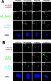
Ser251 phosphorylation of NDEL1 by Aurora-A facilitates centrosomal targeting of NDEL1 and ubiquitin-mediated protein degradation. (A) Replacement experiment of endogenous NDEL1 by wild-type GFP-NDEL1, GFP-NDEL1(S251A), or GFP-NDEL1(S251E) with/without MG132. Wild-type GFP-NDEL1 displayed normal localization. In contrast, GFP-NDEL1(S251A) displayed diffuse distribution throughout the cell cycle, whereas GFP-NDEL1(S251E) displayed more restricted localization at the centrosome in G2 prophase, followed by its disappearance after progression to prophase. This rapid disappearance was prevented by the administration of MG132. Interestingly, the expression of an excess of the GFP-NDEL1(S251E) mutant resulted in multipolar centrosomes rather than their disappearance and the majority of them arrested at prophase. (B) Replacement experiments of endogenous NDEL1 by GFP-NDEL1(tripleS/T→E). GFP-NDEL1(tripleS/T→E) displayed a diffuse distribution pattern like that of GFP-NDEL1(S251A). In addition, a signal was not detected by an anti-phospho-S251 antibody, supporting the interpretation that the phosphorylation by CDK1 inhibited further phosphorylation by Aurora-A. (C) Replacement experiment of endogenous NDEL1 by GFP-NDEL1(tripleS/T→A;S251E) or GFP-NDEL1(tripleS/T→E;S251E). Both mutant NDEL1s displayed centrosomal patterns, but the expression of GFP-NDEL1(tripleS/T→E;S251E) was more unstable and associated with multipolar centrosomes. (D) Expression of GFP-NDEL1(tripleS/T→E;S251E) in wild-type cells. Multipolar centrosomes were also observed in wild-type cells by expression of GFP-NDEL1(tripleS/T→E;S251E), suggesting that this phenotype may be dominant. (E) Western blot using an anti-Ub antibody and an anti-NDEL1 antibody as indicated. Synchronized MEF cells were transfected by expression vectors carrying wild-type GFP-NDEL1, GFP-NDEL1(S251A), or GFP-NDEL1(S251E), and this was followed by immunoprecipitation (IP) at a different time (indicated above panel). Wild-type GFP-NDEL1 was preferentially ubiquitinated at prophase (16 h). Note the decreased ubiquitination of GFP-NDEL1(S251A), whereas enhanced ubiquitination was observed with GFP-NDEL1(S251E). IB, immunoblot; α, anti.
FIG. 3.
FIG. 3.
Ser251 phosphorylation of NDEL1 by Aurora-A facilitates centrosomal targeting of NDEL1 and ubiquitin-mediated protein degradation. (A) Replacement experiment of endogenous NDEL1 by wild-type GFP-NDEL1, GFP-NDEL1(S251A), or GFP-NDEL1(S251E) with/without MG132. Wild-type GFP-NDEL1 displayed normal localization. In contrast, GFP-NDEL1(S251A) displayed diffuse distribution throughout the cell cycle, whereas GFP-NDEL1(S251E) displayed more restricted localization at the centrosome in G2 prophase, followed by its disappearance after progression to prophase. This rapid disappearance was prevented by the administration of MG132. Interestingly, the expression of an excess of the GFP-NDEL1(S251E) mutant resulted in multipolar centrosomes rather than their disappearance and the majority of them arrested at prophase. (B) Replacement experiments of endogenous NDEL1 by GFP-NDEL1(tripleS/T→E). GFP-NDEL1(tripleS/T→E) displayed a diffuse distribution pattern like that of GFP-NDEL1(S251A). In addition, a signal was not detected by an anti-phospho-S251 antibody, supporting the interpretation that the phosphorylation by CDK1 inhibited further phosphorylation by Aurora-A. (C) Replacement experiment of endogenous NDEL1 by GFP-NDEL1(tripleS/T→A;S251E) or GFP-NDEL1(tripleS/T→E;S251E). Both mutant NDEL1s displayed centrosomal patterns, but the expression of GFP-NDEL1(tripleS/T→E;S251E) was more unstable and associated with multipolar centrosomes. (D) Expression of GFP-NDEL1(tripleS/T→E;S251E) in wild-type cells. Multipolar centrosomes were also observed in wild-type cells by expression of GFP-NDEL1(tripleS/T→E;S251E), suggesting that this phenotype may be dominant. (E) Western blot using an anti-Ub antibody and an anti-NDEL1 antibody as indicated. Synchronized MEF cells were transfected by expression vectors carrying wild-type GFP-NDEL1, GFP-NDEL1(S251A), or GFP-NDEL1(S251E), and this was followed by immunoprecipitation (IP) at a different time (indicated above panel). Wild-type GFP-NDEL1 was preferentially ubiquitinated at prophase (16 h). Note the decreased ubiquitination of GFP-NDEL1(S251A), whereas enhanced ubiquitination was observed with GFP-NDEL1(S251E). IB, immunoblot; α, anti.
FIG.4.
FIG.4.
Biochemical and genetic interaction between Aurora-A NDEL1 and TACC3. (A) Coimmunoprecipitation assays. HeLa cells were transfected with expression vectors as indicated. Immunoprecipitation (IP) was carried out using anti-GFP (α-GFP), and immunoblotting (IB) was performed with the anti-NDEL1 or anti-TACC3 antibody. Notice that the mobility of GFP-TACC3 is larger than the expected molecular mass from previous reports (19). (B) Coimmunoprecipitation assays using an anti-NDEL1 antibody. Endogenous NDEL1 clearly binds to TACC3. (C) GST pull-down. In vitro-translated HIS-NDEL1 and HIS-TACC3 were purified and incubated with purified GST-TACC3 and GST-NDEL, respectively, which were immobilized on glutathione-Sepharose 4B beads. Bound proteins were analyzed by Western blotting using an anti-HIS antibody. (D) Examination of phosphorylation and subcellular localization of NDEL1 and TACC3 in Aurora-A-depleted HeLa cells. Aurora-A-depleted HeLa cells exhibited dispersed distribution of NDEL1 and TACC3. The phosphorylation of both proteins was undetectable. (E) Examination of the phosphorylation and subcellular localization of Aurora-A and TACC3 in Ndel1-disrupted MEF cells. TACC3 appeared broadly distributed and lacked phosphorylation, whereas Aurora-A displayed phosphorylation at T288, suggesting that Aurora-A was activated despite the disruption of Ndel1 and that NDEL1 triggers centrosomal targeting of TACC3 and phosphorylation. (F) Examination of phosphorylation and subcellular localization of Aurora-A and NDEL1 in TACC3-depleted HeLa cells. Phosphorylation and subcellular localization of Aurora-A and NDEL1 appeared grossly normal despite the depletion of TACC3. RNAi, RNA interference; −, absence of; +, presence of.
FIG.4.
FIG.4.
Biochemical and genetic interaction between Aurora-A NDEL1 and TACC3. (A) Coimmunoprecipitation assays. HeLa cells were transfected with expression vectors as indicated. Immunoprecipitation (IP) was carried out using anti-GFP (α-GFP), and immunoblotting (IB) was performed with the anti-NDEL1 or anti-TACC3 antibody. Notice that the mobility of GFP-TACC3 is larger than the expected molecular mass from previous reports (19). (B) Coimmunoprecipitation assays using an anti-NDEL1 antibody. Endogenous NDEL1 clearly binds to TACC3. (C) GST pull-down. In vitro-translated HIS-NDEL1 and HIS-TACC3 were purified and incubated with purified GST-TACC3 and GST-NDEL, respectively, which were immobilized on glutathione-Sepharose 4B beads. Bound proteins were analyzed by Western blotting using an anti-HIS antibody. (D) Examination of phosphorylation and subcellular localization of NDEL1 and TACC3 in Aurora-A-depleted HeLa cells. Aurora-A-depleted HeLa cells exhibited dispersed distribution of NDEL1 and TACC3. The phosphorylation of both proteins was undetectable. (E) Examination of the phosphorylation and subcellular localization of Aurora-A and TACC3 in Ndel1-disrupted MEF cells. TACC3 appeared broadly distributed and lacked phosphorylation, whereas Aurora-A displayed phosphorylation at T288, suggesting that Aurora-A was activated despite the disruption of Ndel1 and that NDEL1 triggers centrosomal targeting of TACC3 and phosphorylation. (F) Examination of phosphorylation and subcellular localization of Aurora-A and NDEL1 in TACC3-depleted HeLa cells. Phosphorylation and subcellular localization of Aurora-A and NDEL1 appeared grossly normal despite the depletion of TACC3. RNAi, RNA interference; −, absence of; +, presence of.
FIG.5.
FIG.5.
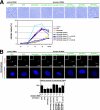
Phosphorylation-mimetic NDEL1 mutant rescued impairment of separation and maturation defect of the centrosome by Aurora-A depletion. (A) To examine whether the expression of phosphorylation-mimetic mutants of NDEL1 support mitotic entry of the HeLa cells in which Aurora-A is depleted by siRNA, we measured the mitotic index of synchronized HeLa cells. Phase-contrast images of cells transfected with control GFP and GFP-Ndel1 constructs and a time course of the mitotic index of cells transfected (shown at the bottom). (B) γ-Tubulin is an essential component for MT nucleation. Centrosomes mature as cells enter mitosis, accumulating γ-tubulin. This occurs concomitantly with an increase in the number of centrosomally organized MTs. We examined the recovery of γ-Tubulin accumulation by expression of mutated NDEL1s under Aurora-A depletion. γ-Tubulin accumulation was estimated by the distribution and intensity of the immunocytochemistry, as indicated. The expression of GFP-NDEL1(S251E), GFP-NDEL1(tripleS/T→A;S251E), and GFP-NDEL1(tripleS/T→E;S251E) improved γ-tubulin accumulation to the centrosome, whereas control GFP, GFP-NDEL1, GFP-NDEL1(S251A), and GFP-NDEL1(tripleS/T→E) did not. The accumulation of γ-tubulin was quantitated by measuring the relative fluorescence intensity of 200-nm circles around the centrosome. Error bars indicate standard deviations. (C) Examination of subcellular localization of TACC3 in Aurora-A-depleted HeLa cells by the expression of mutant Ndel1s. Aurora-A depletion resulted in dispersed localization of TACC3. Exogenous expression of GFP-NDEL1(S251E) restored the centrosomal distribution of TACC3 as indicated. In contrast, the expression of wild-type GFP-NDEL1 or GFP-NDEL1(S251A) failed to restore the impairment of TACC3 localization. (D) At prophase, the separation of centrosomes commenced and moved to opposite poles, associated with a maturation step. HeLa cells that lack Aurora-A displayed an impairment of separation and an attached appearance. The impairment of separation of the centrosomes was efficiently and partially rescued by GFP-NDEL1(S251E) and GFP-NDEL1(tripleS/T→A;S251E), respectively. Control GFP, GFP-NDEL1, GFP-NDEL1(S251A), and GFP-NDEL1(tripleS/T→E) were less efficient in rescuing this phenotype. Note the separation of multiple centrosomes after the transfection of GFP-NDEL1(tripleS/T→E;S251E). Quantitation was carried out by measuring the distance between two centrosomes as shown at the bottom. Error bars indicate standard deviations. RNAi, RNA interference; −, without; +, with.
FIG.5.
FIG.5.
Phosphorylation-mimetic NDEL1 mutant rescued impairment of separation and maturation defect of the centrosome by Aurora-A depletion. (A) To examine whether the expression of phosphorylation-mimetic mutants of NDEL1 support mitotic entry of the HeLa cells in which Aurora-A is depleted by siRNA, we measured the mitotic index of synchronized HeLa cells. Phase-contrast images of cells transfected with control GFP and GFP-Ndel1 constructs and a time course of the mitotic index of cells transfected (shown at the bottom). (B) γ-Tubulin is an essential component for MT nucleation. Centrosomes mature as cells enter mitosis, accumulating γ-tubulin. This occurs concomitantly with an increase in the number of centrosomally organized MTs. We examined the recovery of γ-Tubulin accumulation by expression of mutated NDEL1s under Aurora-A depletion. γ-Tubulin accumulation was estimated by the distribution and intensity of the immunocytochemistry, as indicated. The expression of GFP-NDEL1(S251E), GFP-NDEL1(tripleS/T→A;S251E), and GFP-NDEL1(tripleS/T→E;S251E) improved γ-tubulin accumulation to the centrosome, whereas control GFP, GFP-NDEL1, GFP-NDEL1(S251A), and GFP-NDEL1(tripleS/T→E) did not. The accumulation of γ-tubulin was quantitated by measuring the relative fluorescence intensity of 200-nm circles around the centrosome. Error bars indicate standard deviations. (C) Examination of subcellular localization of TACC3 in Aurora-A-depleted HeLa cells by the expression of mutant Ndel1s. Aurora-A depletion resulted in dispersed localization of TACC3. Exogenous expression of GFP-NDEL1(S251E) restored the centrosomal distribution of TACC3 as indicated. In contrast, the expression of wild-type GFP-NDEL1 or GFP-NDEL1(S251A) failed to restore the impairment of TACC3 localization. (D) At prophase, the separation of centrosomes commenced and moved to opposite poles, associated with a maturation step. HeLa cells that lack Aurora-A displayed an impairment of separation and an attached appearance. The impairment of separation of the centrosomes was efficiently and partially rescued by GFP-NDEL1(S251E) and GFP-NDEL1(tripleS/T→A;S251E), respectively. Control GFP, GFP-NDEL1, GFP-NDEL1(S251A), and GFP-NDEL1(tripleS/T→E) were less efficient in rescuing this phenotype. Note the separation of multiple centrosomes after the transfection of GFP-NDEL1(tripleS/T→E;S251E). Quantitation was carried out by measuring the distance between two centrosomes as shown at the bottom. Error bars indicate standard deviations. RNAi, RNA interference; −, without; +, with.

References

    1. Berdnik, D., and J. A. Knoblich. 2002. Drosophila Aurora-A is required for centrosome maturation and actin-dependent asymmetric protein localization during mitosis. Curr. Biol. 12:640-647. - PubMed
    1. Bischoff, J. R., and G. D. Plowman. 1999. The Aurora/Ipl1p kinase family: regulators of chromosome segregation and cytokinesis. Trends Cell Biol. 9:454-459. - "VSports手机版" PubMed
    1. Cheeseman, I. M., S. Anderson, M. Jwa, E. M. Green, J. Kang, J. R. Yates III, C. S. Chan, D. G. Drubin, and G. Barnes. 2002. Phospho-regulation of kinetochore-microtubule attachments by the Aurora kinase Ipl1p. Cell 111:163-172. - PubMed
    1. Dobyns, W. B. 1987. Developmental aspects of lissencephaly and the lissencephaly syndromes. Birth Defects 23:225-241. - PubMed (VSports手机版)
    1. Dobyns, W. B., O. Reiner, R. Carrozzo, and D. H. Ledbetter. 1993. Lissencephaly: a human brain malformation associated with deletion of the LIS1 gene located at chromosome 17p13. JAMA 23:2838-2842. - PubMed

Publication types

"VSports" MeSH terms