<noscript lang="8tG3B0j"></noscript><ins id="cwqg3t"></ins> Skip to main page content
U.S. flag

An official website of the United States government

Dot gov

The . gov means it’s official. Federal government websites often end in VSports app下载. gov or . mil. Before sharing sensitive information, make sure you’re on a federal government site. .

Https

The site is secure. The https:// ensures that you are connecting to the official website and that any information you provide is encrypted and transmitted securely V体育官网. .

. 2006 Nov;116(11):3042-9.
doi: 10.1172/JCI28746. Epub 2006 Oct 19.

Impaired regulation of NF-kappaB and increased susceptibility to colitis-associated tumorigenesis in CYLD-deficient mice

Affiliations

Impaired regulation of NF-kappaB and increased susceptibility to colitis-associated tumorigenesis in CYLD-deficient mice

VSports注册入口 - Jun Zhang et al. J Clin Invest. 2006 Nov.

Abstract

Cylindromatosis (CYLD) is a deubiquitinating enzyme that is altered in patients with familial cylindromatosis, a condition characterized by numerous benign adnexal tumors. However, the regulatory function of CYLD remains unsettled. Here we show that the development of B cells, T cells, and myeloid cells was unaffected in CYLD-deficient mice, but that the activation of these cells with mediators of innate and adaptive immunity resulted in enhanced NF-kappaB and JNK activity associated with increased TNF receptor-associated factor 2 (TRAF2) and NF-kappaB essential modulator (NEMO) ubiquitination. CYLD-deficient mice were more susceptible to induced colonic inflammation and showed a dramatic increase in the incidence of tumors compared with controls in a colitis-associated cancer model. These results suggest that CYLD limits inflammation and tumorigenesis by regulating ubiquitination in vivo. VSports手机版.


"VSports注册入口" Figures
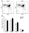
Figure 1
Figure 1. T and B cell development in Cyld–/– mice.
(A) Thymocytes from 4- to 6-week-old mice were analyzed for CD4 and CD8 expression. In each panel 10,000 cells were analyzed. Numbers indicate the percentage of cells in each subset. Data are representative of 4 experiments with 5 mice of each genotype. (B) Spleen populations as determined by flow cytometry from 6- to 8-week-old mice. Data are mean ± SD calculated from 5 mice of each genotype.
Figure 2
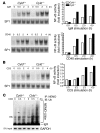
Figure 2. CYLD is a negative regulator of NF-κB activation in B and T cells.
(A) Cellular extracts prepared from splenic B cells stimulated with anti-IgM (10 μg/ml) or CD40 agonist Ab (1 μg/ml) were analyzed by EMSA for NF-κB binding. (B) Cellular extracts prepared from splenic T cells and stimulated with anti-CD3 were analyzed by EMSA for NF-κB binding. Quantification of NF-κB activity in A and B is represented as fold change compared with unstimulated cells (set at 1.0). Ub, ubiquitin. (C) Following T cell stimulation with anti-CD3, NEMO ubiquitination was analyzed by immunoprecipitation of the proteins from denatured cellular lysates followed by immunoblotting with an anti-ubiquitin Ab. NEMO-(Ub)n, polyubiquitinylated NEMO; ns, nonspecific; SP1, specificity protein 1.
Figure 3
Figure 3. Comparison of triggered NF-κB activation and cytokine production in Cyld–/– and wild-type cells.
(A) Peritoneal macrophages were stimulated with TNF-α (500 U/ml), CD40 agonist Ab (1 μg/ml), LPS (2.5 μg/ml), or Pam3CSK4 (150 ng/ml). Cellular extracts were prepared from cells stimulated for the indicated intervals and analyzed by EMSA for NF-κB binding activity. Quantification of NF-κB activity is represented as fold change compared with unstimulated cells (set at 1.0). (B) Peritoneal macrophages were stimulated with LPS (2.5 μg/ml), Pam3CSK4 (150 ng/ml), or CD40 agonist Ab (5 μg/ml) for 24 hours. IFN-γ (10 ng/ml) was added to all stimulation conditions, and the production of IL-6 and TNF-α into culture supernatants was assessed by ELISA. Med, medium. *P < 0.005, Cyld–/– versus wild-type. (C) CYLD overexpression suppresses NF-κB activation by CD40, EDAR, and RANK, but not by TNF-α. 293M cells were transfected with an NF-κB reporter construct either in combination with a CYLD-bearing plasmid or with empty vector as a control. Cells were stimulated with TNF-α, and luciferase activity was measured. To activate other TNFR pathways, vectors bearing CD40, EDAR, and RANK were cotransfected into 293M cells. This led to receptor overexpression and ligand-independent NF-κB activation caused by spontaneous receptor trimerization. Quantitation of NF-κB activity in the presence of CYLD is indicated as fold suppression. *P < 0.005, Cyld–/– versus vector.
Figure 4
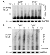
Figure 4. Comparison of TRAF2 and RIP polyubiquitination levels in Cyld–/– and wild-type cells.
(A) Peritoneal macrophages stimulated with TNF-α. TRAF2 ubiquitination was analyzed by immunoprecipitation of the proteins from denatured cellular lysates followed by immunoblotting with an anti-ubiquitin Ab. (B) Lysates prepared from peritoneal macrophages stimulated with TNF-α were immunoprecipitated with an RIP-specific Ab followed by immunoblotting with an anti-ubiquitin Ab.
Figure 5
Figure 5. Comparison of cellular JNK levels in Cyld–/– and wild-type cells.
(A) Lysates prepared from peritoneal macrophages stimulated with LPS or TNF-α were Western blotted with phosphospecific (P-) antibodies recognizing activated JNKs. (B) Lysates prepared from T cells stimulated with anti-CD3 were Western blotted with phosphospecific antibodies recognizing activated JNKs. SAPK, stress-activated protein kinase.
Figure 6
Figure 6. Increased colonic inflammation and tumor development in Cyld–/– mice.
(A) Schematic of the AOM and DSS CAC model. (B) Weight loss during the first cycle of DSS treatment in Cyld–/– and wild-type mice, expressed as percent of the starting weight for each cohort. (C) Histologies of Cyld–/– and wild-type colons, either left untreated or 7 days after the first cycle of DSS treatment. Magnification, ×50. (D) Survival curves of Cyld–/– and wild-type mice (n = 60 per group) injected with AOM followed by DSS. Curves are statistically different (P < 0.001). (E) Incidence and total number of tumors (>0.5 mm) in the colons of Cyld–/– and wild-type mice after administration of AOM followed by DSS. Mice were sacrificed 7 days after the first (left) or the second cycle (right) of DSS treatment. *P < 0.001; Cyld–/– versus wild-type. (F) Typical colon histologies demonstrating broad-based adenocarcinoma in a Cyld–/– section 7 days after the second cycle of DSS treatment. Magnification, ×50.
Figure 7
Figure 7. COX-2 and iNOS immunostaining of tissue sections obtained from wild-type and Cyld–/–mice 7 days after the first, second, and third cycles of DSS treatment.
Numerous COX-2–positive cells were observed in the epithelium, and iNOS-positive cells were observed within broadly based inflammatory infiltrating cells, of the adenomatous tissue of Cyld–/– mice. Arrowheads indicate positively stained cells. Magnification, ×50.

V体育官网 - References

    1. Kovalenko A., et al. The tumour suppressor CYLD negatively regulates NF-kappaB signalling by deubiquitination. Nature. 2003;424:801–805. - PubMed
    1. Trompouki E., et al. CYLD is a deubiquitinating enzyme that negatively regulates NF-kappaB activation by TNFR family members. Nature. 2003;424:793–796. - PubMed
    1. Brummelkamp T.R., Nijman S.M., Dirac A.M., Bernards R. Loss of the cylindromatosis tumour suppressor inhibits apoptosis by activating NF-kappaB. Nature. 2003;424:797–801. - PubMed
    1. Makarova K.S., Aravind L., Koonin E.V. A novel superfamily of predicted cysteine proteases from eukaryotes, viruses and Chlamydia pneumoniae. Trends Biochem. Sci. 2000;25:50–52. - VSports app下载 - PubMed
    1. Brooke H. Epithelioma adenoides cysticum. Br. J. Dermatol. 1892;4:269–287.

MeSH terms