Skip to main page content
U.S. flag

An official website of the United States government

Dot gov

The . gov means it’s official. Federal government websites often end in VSports app下载. gov or . mil. Before sharing sensitive information, make sure you’re on a federal government site. .

Https

The site is secure. The https:// ensures that you are connecting to the official website and that any information you provide is encrypted and transmitted securely V体育官网. .

. 2006 Oct;116(10):2707-16.
doi: 10.1172/JCI25546. Epub 2006 Sep 14.

Mll partial tandem duplication induces aberrant Hox expression in vivo via specific epigenetic alterations

Affiliations

VSports注册入口 - Mll partial tandem duplication induces aberrant Hox expression in vivo via specific epigenetic alterations

Adrienne M Dorrance et al. J Clin Invest. 2006 Oct.

Abstract

We previously identified a rearrangement of mixed-lineage leukemia (MLL) gene (also known as ALL-1, HRX, and HTRX1), consisting of an in-frame partial tandem duplication (PTD) of exons 5 through 11 in the absence of a partner gene, occurring in approximately 4%-7% of patients with acute myeloid leukemia (AML) and normal cytogenetics, and associated with a poor prognosis. The mechanism by which the MLL PTD contributes to aberrant hematopoiesis and/or leukemia is unknown. To examine this, we generated a mouse knockin model in which exons 5 through 11 of the murine Mll gene were targeted to intron 4 of the endogenous Mll locus. Mll(PTD/WT) mice exhibit an alteration in the boundaries of normal homeobox (Hox) gene expression during embryogenesis, resulting in axial skeletal defects and increased numbers of hematopoietic progenitor cells. Mll(PTD/WT) mice overexpress Hoxa7, Hoxa9, and Hoxa10 in spleen, BM, and blood. An increase in histone H3/H4 acetylation and histone H3 lysine 4 (Lys4) methylation within the Hoxa7 and Hoxa9 promoters provides an epigenetic mechanism by which this overexpression occurs in vivo and an etiologic role for MLL PTD gain of function in the genesis of AML. VSports手机版.


VSports在线直播 - Figures

Figure 1
Figure 1. Generation of MllPTD/WT mice.
The backbone of the targeting vector is ploxPneo1 and contains a genomic clone spanning Mll intron 4 through intron 11. Linearization of the vector at a unique SfiI site within intron 4 allows for recombination at the germline intron 4 of Mll. A plasmid vector expressing Cre recombinase from a CMV promoter (pCre) was used to excise the neo cassette.
Figure 2
Figure 2. Germline transmission and verification of correct targeting in mice heterozygous for the Mll PTD.
(A) Southern blot analysis using high molecular-weight DNA from spleens, digested with NdeI and hybridized to a probe that spans intron 4 and exon 5 of Mll (i4e5 in Figure 1). This generates a single WT band at 18 kb in MllWT/WT mice, 2 bands representing 1 WT allele at 18 kb, and a band at 40 kb representing the rearranged allele in MllPTD/WT mice. (B) The Mll PTD fusion transcript was amplified using an upstream primer from Mll exon 11 and a downstream primer from Mll exon 6 amplifying a single 335-bp unique fusion transcript in the MllPTD/WT mouse (+) and is absent in the sample from the MllWT/WT mouse (–). Sequencing of these PCR products from the MllPTD/WT mice verified the presence of the exon 11–exon 5 fusion site. The 1-kb+ ladder was used to determine amplification size.
Figure 3
Figure 3. Mll WT and Mll PTD transcripts.
Schematic demonstrating the real-time RT-PCR strategy for detection and absolute quantification of the Mll WT and Mll PTD transcripts in bone marrow and spleen of Mll PTD mice. The predicted Mll PTD and Mll WT allele–derived transcripts are shown, with the tandemly duplicated exons 5–11 present in the PTD transcript denoted with light gray boxes. Shown below the transcripts are sites for PCR primers and fluorogenic probes designed to amplify either the unique exon 11–exon 5 fusion (open rectangle) or exons 14–15, whose junction is common to the Mll WT and Mll PTD transcripts (filled rectangle).
Figure 4
Figure 4. Skeletal analysis of MllPTD/WT mice at 20 weeks of age.
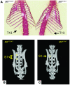
(A) Alizarin red staining of MllPTD/WT (left) and MllWT/WT (right) mice shows a rudimentary/missing thirteenth rib, indicative of a T13 → L1 transformation (penetrance of 70% in MllPTD/WT mice, n = 13). MicroCT images of the sacral spine in a (B) MllPTD/WT mouse and a (C) MllWT/WT mouse in 3D. These images are viewed from the dorsal side and illustrate the duplication of the S1 vertebral body, indicative of an S2→S1 transformation in the MllPTD/WT mice (penetrance of 63%, n = 11).
Figure 5
Figure 5. In situ hybridizations for Hoxa9 on E12.5 embryos.
(A) MllWT/WT and (B) MllPTD/WT embryos hybridized with a digoxygenin-labeled probe for Hoxa9. Results demonstrate a notable ventral shift of the Hoxa9 expression boundary within the paraxial mesoderm of the MllPTD/WT embryo compared with the MllWT/WT littermate control (see inset for each figure). This figure represents 1 of 3 comparable hybridizations.
Figure 6
Figure 6. Evaluation of progenitor populations in MllPTD/WT splenocytes compared with MllWT/WT sex-matched littermate controls.
(A) Results from CFU assays to assess progenitors of erythroid (BFU-E), granulocytic, erythroid, monocytic, megakaryocytic (GEMM-CFU), and granulocyte, macrophage (GM-CFU) lineages show significantly increased CFUs derived from MllPTD/WT versus MllWT/WT splenocytes. *P = 0.03; **P = 0.0025. Error bars indicate SD. Splenocytes from (B) MllWT/WT and (C) MllPTD/WT mice (n = 8 mice per genotype) show increased surface density expression of the erythroid marker Ter119 in MllPTD/WT mice. Ter PE, PE fluorochrome–conjugated Ter119. Secondary splenic colonies from (D) MllWT/WT, (E) MllAf9/WT, and (F) MllPTD/WT mice (n = 6 mice per genotype, plated in duplicate). The numbers of secondary colonies per plate (~230) were similar between genotypes. The number of cells per colony was dramatically increased in MllPTD/WT mice compared with MllWT/WT or MllAf9/WT mice. (GI) Primary CFU progenitor cells harvested after 14 days and maintained in liquid cultures for 18 days, supplemented with SCF, IL-3, and IL-6, and in the presence of BrdU for the last 4 days. An anti-BrdU antibody gated on viable cells demonstrates a smaller fraction of MllWT/WT cells proliferating (G) compared with MllPTD/WT cells (H). Assessment of programmed cell death (I, upper right and lower right quadrants) revealed a sizeable fraction (~50%) of MllPTD/WT progenitors undergoing apoptosis during expansion. While a significant fraction of MllPTD/WT cells were proliferating, approximately 50% were undergoing apoptosis. In 2 additional experiments, the MllWT/WT cells had already died by this time point in culture (see Table 3), precluding a statistical comparison of these results in GI.
Figure 7
Figure 7. HoxA gene expression levels.
(AC) Quantification of Hoxa7, Hoxa9, and Hoxa10 expression in hematopoietic tissues of MllWT/WT and MllPTD/WT mice. Cells were isolated from (A) BM, (B) spleen, and (C) blood from both MllWT/WT and MllPTD/WT mice (n = 10 mice per genotype). RNA and then cDNA were prepared and quantified by real-time RT-PCR. MllPTD/WT mice showed increased expression of each of these HoxA genes in all 3 tissues compared with sex-matched littermate MllWT/WT mice. (D) Equal numbers of each sorted BM population expressing Gr-1, CD11b, CD3, B220, or Ter119 from MllPTD/WT mice and MllWT/WT mice were processed for RNA and then cDNA. Absolute quantification of Hoxa9 expression was then determined by real-time RT-PCR and demonstrated that MllPTD/WT cells have increased Hoxa9 expression within each population compared with the equivalent number of MllWT/WT cells, indicating that Hoxa9 transcript levels are overexpressed on a per cell basis (values shown represent mean of n = 3 mice per genotype ± SD; P < 0.03).
Figure 8
Figure 8. Assessment of Mll target gene Hoxa9 acetylation and methylation of histones assessed by ChIP using (A) bone marrow cells and (B) splenocytes derived from MllPTD/WT and MllWT/WT mice.
Results show an increase at the Hoxa9 promoter in histone H3/H4 acetylation as well as histone H3 Lys4 methylation and a corresponding decrease in histone H3 Lys9 methylation in MllPTD/WT bone marrow and spleen compared with the same MllWT/WT tissues. Results are representative of 3 independent experiments. Similar results were obtained at the Hoxa7 promoter but were not seen at the Hoxa10 promoter (not shown). (C) BM cells and (D) splenocytes derived from MllPTD/WT, MllWT/–, and MllWT/WT mice. Absolute quantification by SYBR green real-time PCR shows a significant increase at the Hoxa9 promoter in histone H3/H4 acetylation as well as histone H3 Lys4 methylation in MllPTD/WT BM and spleen compared with the same MllWT/– and MllWT/WT tissues (n = 3 mice per genotype; P < 0.05). There was no significant difference between MllWT/– and MllWT/WT tissues.

References

    1. Yu B.D., Hess J.L., Horning S.E., Brown G.A., Korsmeyer S.J. Altered Hox expression and segmental identity in Mll-mutant mice. Nature. 1995;378:505–508. - PubMed
    1. Zeleznik-Le N.J., Harden A.M., Rowley J.D. 11q23 translocations split the “AT hook” cruciform DNA binding region and the transcriptional repression domain from the activation domain of the mixed-lineage leukemia (MLL) gene. Proc. Natl. Acad. Sci. U. S. A. 1994;91:10610–10614. - PMC - PubMed
    1. Caligiuri M.A., et al. Molecular rearrangement of the ALL-1 gene in acute myeloid leukemia without cytogenetic evidence of 11q23 chromosomal translocations. Cancer Res. 1994;54:370–373. - "VSports手机版" PubMed
    1. Caligiuri M., et al. Rearrangement of ALL1 (MLL) in acute myeloid leukemia with normal cytogenetics. . Cancer Res. 1998;58:55–59. - PubMed
    1. Dohner K., et al. Prognostic significance of partial tandem duplications of the MLL gene in adult patients 16 to 60 years old with acute myeloid leukemia and normal cytogenetics: a study of the Acute Myeloid Leukemia Study Group Ulm. J. Clin. Oncol. 2002;20:3254–3261. - "V体育安卓版" PubMed

"V体育安卓版" MeSH terms

LinkOut - more resources