Skip to main page content
U.S. flag

An official website of the United States government

Dot gov

The . gov means it’s official. Federal government websites often end in . gov or . mil. Before sharing sensitive information, make sure you’re on a federal government site VSports app下载. .

Https

The site is secure. The https:// ensures that you are connecting to the official website and that any information you provide is encrypted and transmitted securely V体育官网. .

. 2006 Sep;47(9):2028-41.
doi: 10.1194/jlr.M600177-JLR200. Epub 2006 Jun 21.

"V体育2025版" Regulation of hepatic fatty acid elongase and desaturase expression in diabetes and obesity

Affiliations

V体育安卓版 - Regulation of hepatic fatty acid elongase and desaturase expression in diabetes and obesity

Yun Wang et al. J Lipid Res. 2006 Sep.

"VSports" Erratum in

  • J Lipid Res. 2006 Oct;47(10):2353. Nair, Muraleedharan [corrected to Nair, Muraleedharan G]; Peters, Jeffery M [corrected to Peters, Jeffrey M]; Busik, Julia [corrected to Busik, Julia V]

"VSports" Abstract

Fatty acid elongases and desaturases play an important role in hepatic and whole body lipid composition. We examined the role that key transcription factors played in the control of hepatic elongase and desaturase expression. Studies with peroxisome proliferator-activated receptor alpha (PPARalpha)-deficient mice establish that PPARalpha was required for WY14643-mediated induction of fatty acid elongase-5 (Elovl-5), Elovl-6, and all three desaturases [Delta(5) desaturase (Delta(5)D), Delta(6)D, and Delta(9)D]. Increased nuclear sterol-regulatory element binding protein-1 (SREBP-1) correlated with enhanced expression of Elovl-6, Delta(5)D, Delta(6)D, and Delta(9)D. Only Delta(9)D was also regulated independently by liver X receptor (LXR) agonist. Glucose induction of l-type pyruvate kinase, Delta(9)D, and Elovl-6 expression required the carbohydrate-regulatory element binding protein/MAX-like factor X (ChREBP/MLX) heterodimer. Suppression of Elovl-6 and Delta(9)D expression in livers of streptozotocin-induced diabetic rats and high fat-fed glucose-intolerant mice correlated with low levels of nuclear SREBP-1 VSports手机版. In leptin-deficient obese mice (Lep(ob/ob)), increased SREBP-1 and MLX nuclear content correlated with the induction of Elovl-5, Elovl-6, and Delta(9)D expression and the massive accumulation of monounsaturated fatty acids (18:1,n-7 and 18:1,n-9) in neutral lipids. Diabetes- and obesity-induced changes in hepatic lipid composition correlated with changes in elongase and desaturase expression. In conclusion, these studies establish a role for PPARalpha, LXR, SREBP-1, ChREBP, and MLX in the control of hepatic fatty acid elongase and desaturase expression and lipid composition. .


Figures
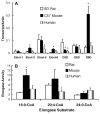
Fig. 1
Fig. 1
Fatty acid elongase and desaturase expression in rat, mouse, and human liver. A: Livers from male Sprague-Dawley (SD) rats (2–3 months of age), C57BL/6 mice (2–4 months of age), and female humans (44–54 years of age) were used for RNA extraction and measurement of elongase and desaturase expression as described in Materials and Methods. Rats and mice were maintained on chow (Teklad) diets ad libitum. Human livers were obtained from the National Disease Research Interchange. mRNA abundance for each elongase and desaturase was measured by quantitative real-time (qRT) PCR. Results are expressed relative to an internal standard, β-actin (transcript/actin) (means ± SD; n = 4 for rat and mouse liver, n = 3 for human liver). B: Rat, mouse, and human liver was extracted for microsomes to assay elongase activity using three separate substrates, 16:0-CoA, 20:4-CoA, and 24:0-CoA. Results are expressed as elongase activity (nmol [14C]malonyl-CoA assimilated into fatty acids/mg protein) (means ± SD; n = 4 for rat and mouse liver, n = 3 for human liver). Δ5D, Δ5 desaturase; Elovl-1, fatty acid elongase-1. * P ≤ 0.05 versus rat liver by ANOVA.
Fig. 2
Fig. 2
Role of peroxisome proliferator-activated receptor α (PPARα) in the control of hepatic elongase and desaturase expression. Homozygous wild-type (+/+) and PPARα-null (−/−) mice on a Sv/129 genetic background (19, 20) were fed either a control diet or one containing WY14643 (50 or 500 mg/kg diet; Bioserv, Piscataway, NJ) for 1 week. Liver RNA was extracted and used as a template for qRT-PCR. Results are reported as fold change in mRNA abundance (transcript/cyclophilin) for each enzyme (means ± SD; n = 4). * P ≤ 0.05 by Student’s t-test versus the wild type (+/+) on a control diet.
Fig. 3
Fig. 3
Effect of insulin and liver X receptor (LXR) agonist on fatty acid elongase and desaturase expression in rat primary hepatocytes. Rat primary hepatocytes were maintained in Williams E medium containing 10 mM lactate and 10 nM dexamethasone with no insulin overnight. The next day, cells were switched to Williams E medium containing 25 mM glucose and 10 nM dexamethasone [vehicle (Veh)] in the presence or absence of insulin (Ins; 1 μM) or the LXR agonist T0901317 (T1317; Cayman Chemical Co., Ann Arbor, MI) (1 μM). After 24 h of treatment, cells were harvested for RNA extraction and the measurement of nuclear sterol-regulatory element binding protein-1 (SREBP-1) and SREBP-2 by immunoblot analysis (inset; duplicate samples/treatment). Elongase and desaturase mRNA abundance was quantified by qRT-PCR. Results are expressed as fold change in mRNA versus vehicle (transcript/ cyclophilin) (means ± SD; n = 4). Results are representative of two separate studies. * P ≤ 0.05 versus vehicle by ANOVA.
Fig. 4
Fig. 4
Effect of overexpressed nuclear SREBP-1c on elongase and desaturase expression. Primary rat hepatocytes in Williams E medium + 10 mM lactate + 10 nM dexamethasone but no insulin were infected with a recombinant adenovirus expressing the nuclear form of SREBP-1c under the control of doxycycline (25). Cells were switched to Williams E medium + 25 mM glucose with no insulin in the absence or presence of doxycycline (250 ng/ml). Cells were harvested 24 h afterward for RNA extraction and measurement of elongase and desaturase mRNA abundance by qRT-PCR (transcript/ cyclophilin). Endogenous SREBP-1 expression was quantified by Northern analysis. Results are represented as fold induction by SREBP-1c (means ± SD; n = 4). Results are representative of two separate studies. * P > 0.05 versus vehicle by ANOVA.
Fig. 5
Fig. 5
Effect of glucose on elongase and desaturase expression. A: Primary rat hepatocytes in Williams E medium + 10 mM lactate + dexamethasone + insulin were switched to Williams E medium + 25 mM glucose + dexamethasone + insulin. After 24 h of treatment, cells were harvested for extraction of nuclear proteins and RNA. The nuclear abundance of carbohydrate-regulatory element binding protein (ChREBP) and MAX-like factor X (MLX) was measured by immunoblotting at the times indicated (22). B: L-type pyruvate kinase (L-PK), Elovl-6, and Δ9D mRNA abundance was quantified by qRT-PCR (transcript/ cyclophilin). Results are expressed as fold induction by glucose (means ± SD; n = 4). C: In a second experiment, primary hepatocytes in Williams E medium + 10 mM lactate + dexamethasone + insulin remained uninfected (white bars) or were infected with recombinant adenovirus expressing luciferase (Ad-Luc; black bars) or dominant negative MLX (Ad-dnMLX; gray bars). After 24 h, cells were switched to Williams E medium + 25 mM glucose + dexamethasone + insulin. Twenty-four hours later, cells were harvested for RNA extraction and measurement of L-PK, Elovl-6, and Δ9D mRNA by qRT-PCR (transcript/cyclophilin). Results are represented as relative mRNA abundance relative to glucose-treated cells (means ± SD; n = 4). Results are representative of two separate studies. * P < 0.001 versus glucose-treated cells by ANOVA.
Fig. 6
Fig. 6
Substrate specificity of hepatic fatty acid elongases. Ad-Luc, Elovl-2, Elovl-5, and Elovl-6 were used to infect primary hepatocytes (5 plaque-forming units/cell). After 24 h, cells were harvested for elongase activity using various fatty acid substrates (see Materials and Methods). Results are expressed as elongase activity (nmol [14C]malonyl-CoA assimilated into fatty acid/mg protein) (means ± SD; n = 3). Results are representative of two separate studies.
Fig. 7
Fig. 7
Effects of insulin and WY14643 on monounsaturated fatty acid synthesis. A: The pathway for conversion of 16:0 to 16:1,n-7, 18:1,n-7, and 18:1,n-9 by Elovl-5, Elovl-6, and Δ9D. B–D: Primary rat hepatocytes in Williams E + dexamethasone + 10 mM lactate overnight were maintained in the same medium or switched to Williams E + dexamethasone + 25 mM glucose in the absence or presence of insulin (1 μM) or WY14643 (50 μM). Cells received 100 μM [14C]16:0 + 20 μM BSA for 24 h. After 24 h of treatment, cells were extracted for total lipid and the lipid was saponified. The distribution of 14C among 16:0, 16:1,n-7, 18:0, 18:1,n-7 and 18:1,n-9 was quantified by reverse-phase HPLC and a flow-through β-scintillation counter. Technical limitations preclude resolution of 18:1,n-7 and 18:1,n-9 by reverse-phase HPLC. As such, the results are reported as 18:1. Results are reported as percentage conversion of [14C]16:0 to [14C]16:1 (B), percentage conversion of [14C]16:0 to [14C]18:1 (C), and ratio of [14C]18:1 to [14C]16:1 (elongation index) (D). Results are means of duplicate samples and are representative of two separate studies.
Fig. 8
Fig. 8
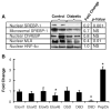
Effect of streptozotocin-induced diabetes on hepatic elongase and desaturase expression. Male Sprague-Dawley rats were made diabetic using streptozotocin as described in Materials and Methods. Livers from control and diabetic animals were used for the isolation of nuclear and microsomal proteins for immunoblotting (A) and RNA extraction for qRT-PCR (B). A: Effect of diabetes on SREBP-1 (microsomal and nuclear), nuclear ChREBP, MLX, and hepatic nuclear factor-4 (HNF-4α) abundance. Protein levels were measured by immunoblot analysis (see Materials and Methods). Duplicate samples for each treatment are shown. The effect of diabetes on the abundance of these proteins was quantified and expressed as fold change (means; n = 5) induced by diabetes. Statistical significance (P) was assessed by Student’s t-test. B: Effect of diabetes on elongase, desaturase, and phosphoenolpyruvate carboxykinase (PepCk) expression. Transcripts encoding fatty acid elongases, fatty acid desaturases, and cyclophilin were quantified by qRT-PCR, whereas phosphoenolpyruvate kinase was quantified by Northern blot analysis. Results are expressed as fold change in expression (transcript/cyclophilin) induced by diabetes (n = 5/group). * P < 0.05 versus control by Student’s t-test.
Fig. 9
Fig. 9
Effect of high-fat diets on hepatic elongase and desaturase expression. Male C57BL/6 mice were fed lard diets [10% calories (low fat) or 60% calories (high fat) as lard] for 10 weeks as described in Materials and Methods (48). A: After 9 weeks on the low- and high-fat diets, mice were assessed for glucose tolerance (see Materials and Methods). Blood glucose was measured at the times indicated. Results are expressed as blood glucose (mg/dl) (means ± SD; n = 8 in each group). B: Effect of dietary fat on SREBP (microsomal and nuclear), ChREBP, MLX, and HNF-4α. After 10 weeks on the diet, livers were recovered for nuclear and microsomal protein and RNA extraction as described for Fig. 8. Nuclear and microsomal proteins were measured as described in Materials and Methods. Representative immunoblots from duplicate samples for each treatment are shown. The effect of dietary fat on the abundance of these proteins was quantified and expressed as fold change (means; n = 4 animals/group) induced by the high-fat diet. Statistical significance (P) was assessed by Student’s t-test. C: Effect of dietary fat on elongase and desaturase expression. RNA was extracted and used for qRT-PCR analysis of elongase and desaturase expression. Results are expressed as fold change (transcript/cyclophilin) (means ± SD; n = 8). * P ≤ 0.05. D: Effect of dietary fat on elongase activity. Hepatic microsomal preparations were used for fatty acid elongase assays (see Materials and Methods). Results are expressed as elongase activity (nmol [14C]malonyl-CoA assimilated into fatty acids/mg protein) (means ± SD; n = 8). * P ≤ 0.05 versus 10% calories as fat by Student’s t-test.
Fig. 10
Fig. 10
Effect of leptin deficiency on hepatic elongase and desaturase expression. Male lean (C57BL/6J-Lepob/+) and obese (C57BL/6J-Lepob/ob) mice (B6.V-Lep ob/J, No. 000632; Jackson Laboratories) were maintained on a Harlan-Teklad laboratory chow (No. 8640) diet and water ad libitum. Livers from these mice were used for the isolation of nuclear and microsomal protein for immunoblotting (A) and RNA extraction for qRT-PCR (B). A: Effect of obesity on SREBP-1 (microsomal and nuclear) and nuclear ChREBP, MLX, and HNF-4α abundance. Protein levels were measured by immunoblot analysis (see Materials and Methods). Triplicate samples for each phenotype are shown. The effect of leptin deficiency on the abundance of these proteins was quantified and expressed as fold change (means; n = 4) induced by leptin deficiency. Statistical significance (P) was assessed by Student’s t-test. B: Effect of leptin deficiency on elongase and desaturase expression. RNA was extracted and used for qRT-PCR analysis of elongase and desaturase expression. Results are expressed as fold change (transcript/cyclophilin) (means ± SD; n = 4). * P < 0.001 versus lean by Student’s t-test. C: Effect of leptin deficiency on elongase activity. Hepatic microsomal preparations were used for fatty acid elongase assays (see Materials and Methods). Results are expressed as elongase activity (nmol [14C]malonyl-CoA assimilated into fatty acids/mg protein) (means ± SD; n = 4). * P ≤ 0.01 versus lean by Student’s t-test. D: Effect of leptin deficiency on hepatic lipid composition. Total lipids were extracted and saponified; fatty acid levels were quantified by reverse-phase HPLC (see Materials and Methods). Results are expressed as fatty acid mol% (means ± SD; n = 4/group). * P ≤ 0.01 versus lean animals by Student’s t-test.

References

    1. Jump DB, Clarke SD. Regulation of gene expression by dietary fat. Annu Rev Nutr. 1999;19:63–90. - PubMed
    1. Jump DB. Fatty acid regulation of gene transcription. Crit Rev Clin Lab Sci. 2004;41:41–78. - "V体育官网" PubMed
    1. Hillgartner FB, Salati LM, Goodridge AG. Physiological and molecular mechanisms involved in nutritional regulation of fatty acid synthesis. Physiol Rev. 1995;75:47–76. - PubMed
    1. Sprecher H. Metabolism of highly unsaturated n-3 and n-6 fatty acids. Biochim Biophys Acta. 2000;1486:219–231. - PubMed
    1. Pawar A, Xu J, Jerks E, Mangelsdorf DJ, Jump DB. Fatty acid regulation of liver X receptors (LXR) and peroxisome proliferator-activated receptor α (PPARα) in HEK293 cells. J Biol Chem. 2002;277:39243–39250. - PubMed

Publication types

MeSH terms

Substances