Skip to main page content
U.S. flag

An official website of the United States government

Dot gov

The VSports app下载. gov means it’s official. Federal government websites often end in . gov or . mil. Before sharing sensitive information, make sure you’re on a federal government site. .

Https

The site is secure. The https:// ensures that you are connecting to the official website and that any information you provide is encrypted and transmitted securely. V体育官网.

. 2006 May;2(5):e45.
doi: 10.1371/journal.ppat.0020045. Epub 2006 May 19.

Recruitment of BAD by the Chlamydia trachomatis vacuole correlates with host-cell survival (VSports)

Affiliations

Recruitment of BAD by the Chlamydia trachomatis vacuole correlates with host-cell survival

Philippe Verbeke et al. PLoS Pathog. 2006 May.

Abstract

Chlamydiae replicate intracellularly in a vacuole called an inclusion. Chlamydial-infected host cells are protected from mitochondrion-dependent apoptosis, partly due to degradation of BH3-only proteins. The host-cell adapter protein 14-3-3beta can interact with host-cell apoptotic signaling pathways in a phosphorylation-dependent manner. In Chlamydia trachomatis-infected cells, 14-3-3beta co-localizes to the inclusion via direct interaction with a C. trachomatis-encoded inclusion membrane protein. We therefore explored the possibility that the phosphatidylinositol-3 kinase (PI3K) pathway may contribute to resistance of infected cells to apoptosis VSports手机版. We found that inhibition of PI3K renders C. trachomatis-infected cells sensitive to staurosporine-induced apoptosis, which is accompanied by mitochondrial cytochrome c release. 14-3-3beta does not associate with the Chlamydia pneumoniae inclusion, and inhibition of PI3K does not affect protection against apoptosis of C. pneumoniae-infected cells. In C. trachomatis-infected cells, the PI3K pathway activates AKT/protein kinase B, which leads to maintenance of the pro-apoptotic protein BAD in a phosphorylated state. Phosphorylated BAD is sequestered via 14-3-3beta to the inclusion, but it is released when PI3K is inhibited. Depletion of AKT through short-interfering RNA reverses the resistance to apoptosis of C. trachomatis-infected cells. BAD phosphorylation is not maintained and it is not recruited to the inclusion of Chlamydia muridarum, which protects poorly against apoptosis. Thus, sequestration of BAD away from mitochondria provides C. trachomatis with a mechanism to protect the host cell from apoptosis via the interaction of a C. trachomatis-encoded inclusion protein with a host-cell phosphoserine-binding protein. .


Conflict of interest statement

Competing interests. The authors have declared that no competing interests exist V体育安卓版.

Figures

Figure 1
Figure 1. Signal-Transduction Pathways Involved in Resistance of C. trachomatis-Infected Cells against Apoptosis
All cells were infected with C. trachomatis at an MOI of 1.0 for 32 h and then treated overnight with 2 μM STS. In uninfected cells that had been treated with STS under the same conditions, 83 ± 10% of the cells had apoptotic nuclei. Inhibitors were added 24 h before STS treatment, as indicated in the inset. The inhibitor concentrations (and targeted mediators) were: 50 μM LY294002 (PI3K), 10 μM SB203580 (P38), 10 μM U0126 (MEK5), 10 μM SP600125 (JNK1/2), 5 nM rapamycin (mTOR), and 50 μM KNK437 (heat shock proteins). The “control (no STS)” contained infected cells, while “control w/STS” contained infected cells that were incubated with buffer without inhibitor and then treated with STS. DNA was revealed by Hoechst staining, and values are expressed as the % of infected cells that had apoptotic nuclei. The average and SD were calculated from the values obtained from at least 50 microscope fields. The experiment was performed on three separate days, and results from a representative experiment are shown.
Figure 2
Figure 2. Cytochrome c Release from C. trachomatis-Infected Cells Treated with the PI3K Inhibitor
Cells were infected with C. trachomatis at an MOI of 1.0 for 26 h (B) or mock-infected (A), and then incubated with control buffer or 50 μM LY294002 (LY) or control buffer for 6 h before treating with 2 μM STS overnight. Cells were then collected for cytofluorimetry, permeabilized with detergent, and incubated with anti-cytochrome c antibody, as described in Materials and Methods. Cytochrome c was released from uninfected cells that were treated with STS (A), but was retained in infected cells treated with STS (B). However, cytochrome c was released in infected cells that had been pre-incubated with LY294002 before STS treatment (B). One experiment of two representative experiments performed on separate days is shown.
Figure 3
Figure 3. AKT Phosphorylation in Cells during Apoptosis of C. trachomatis-Infected Cells
Cells were infected with C. trachomatis at an MOI of 1.0 for 24 or 48 h, or mock-infected, and then incubated with 50 μM LY294002 (LY) or control buffer for 6 h before treating with control buffer or 2 μM STS overnight. Cells were then collected for Western immunoblotting and analyzed for phosphorylation of AKT on residue Ser473, as described in Materials and Methods. STS treatment by itself did not affect AKT Ser473 phosphorylation in infected cells, but LY294002 caused AKT Ser473 to become partially dephosphorylated. This residue became dephosphorylated almost completely when LY294002 was combined with STS in infected cells. One experiment of two representative experiments performed on separate days is shown.
Figure 4
Figure 4. Effect of Inhibition of AKT Expression on Apoptosis of C. trachomatis-Infected Cells
Cells were transfected with AKT siRNA (+) or control reagents (−) for 8 h, infected with C. trachomatis at an MOI of 1.0 for 32 h, and then incubated with 2 μM STS or control buffer overnight. The efficiency of transfection with AKT siRNA in epithelial cells was monitored by co-transfecting with fluorescein-labeled irrelevant siRNA, which showed that approximately 80% of cells were transfected during the experiments (unpublished data). DNA was revealed with Hoechst and Chlamydia with anti-Chlamydia antibody. The extent of nuclear condensation was measured only in cells that contained Chlamydia vacuoles. No nuclear condensation was observed in infected cells that expressed normal levels of AKT, but STS led to nuclear condensation in infected cells that had been transfected with AKT siRNA. One experiment of two representative experiments performed on separate days is shown. Inset: Inhibition of AKT expression in epithelial cells by RNA interference. Cells were transfected with siRNA for AKT or controls. After 48 h, the cells were harvested and AKT protein levels were measured by Western immunoblotting. The control reagents did not affect AKT expression, and AKT siRNA had no effect on the expression of another protein, the P42 MAP kinase.
Figure 5
Figure 5. Time Course of BAD Phosphorylation during C. trachomatis or C. muridarum Infection
(A) Epithelial cells were transfected with a BAD expression plasmid prior to infection, as described in Materials and Methods. Cells over-expressing BAD were infected with C. trachomatis at an MOI of 1.0 for the indicated times. Cells were then collected for Western immunoblotting and analyzed for AKT phosphorylation (top row), total AKT protein (second row), total BAD protein (third row), phosphorylation of BAD on residue Ser136 (fourth row), or actin (bottom row), as described in Materials and Methods. Chlamydia infection at this MOI led to a decrease in the level of total BAD protein after 32 h of infection. For AKT residue Ser473, there was a low level of phosphorylation in uninfected cells (time 0), but there was a noticeable increase in phosphorylation for both AKT and BAD after 16 h of infection. Phosphorylation levels decreased after 32 h of infection for both BAD and AKT. Chlamydia infection had no effect on actin protein levels (bottom row). One experiment of three representative experiments performed on separate days is shown. (B) Cells expressing endogenous levels of BAD were infected with C. trachomatis at an MOI of 1.0 for the indicated times. Cells were then collected for Western immunoblotting and analyzed for phosphorylation of BAD on residue Ser136 (top row) or actin (bottom row). The time course of BAD phosphorylation in cells expressing only endogenous BAD was similar to the time course in cells that had been transfected with the BAD expression plasmid (A). One experiment of two representative experiments performed on separate days is shown. (C) Cells over-expressing BAD were infected with C. muridarum at an MOI of 1.0 for the indicated times. Cells were then collected for Western immunoblotting and analyzed for AKT phosphorylation (top row), total AKT protein (second row), total BAD protein (third row), phosphorylation of BAD on residue Ser136 (fourth row), or actin (bottom row). Chlamydia infection at this low MOI led to a decrease in the level of total BAD protein after 32 h of infection. BAD was phosphorylated significantly within 5 h of infection, but phosphorylation levels decreased by 8 h post-infection and were not measurable after 1 d of infection. One experiment of three representative experiments performed on separate days is shown.
Figure 6
Figure 6. Effect of PI3K on BAD Phosphorylation during C. trachomatis Infection
Cells over-expressing BAD were infected with C. trachomatis at an MOI of 1.0 for 26 h or mock-infected, and then incubated with 50 μM LY294002 (LY) or control buffer for 6 h before treating with 2 μM STS overnight. Cells were then collected for Western immunoblotting and analyzed for total BAD protein (top row), phosphorylation of BAD on residue Ser136 (middle row), or total P42 MAP kinase protein (bottom row), as described in Materials and Methods. C. trachomatis infection led to a decrease in the level of BAD, which was further decreased by STS treatment. Pre-incubation with LY294002 did not further alter the level of total BAD (top row). STS-treatment of uninfected cells caused complete dephosphorylation of BAD, but BAD remained phosphorylated after STS-treatment of infected cells (middle row). However, pre-incubation of infected cells with LY294002 before treatment with STS resulted in complete dephosphorylation of BAD (middle row). Infection had no effect on total P42 MAP kinase protein levels (bottom row). One experiment of three representative experiments performed on separate days is shown.
Figure 7
Figure 7. Localization of BAD and Phosphorylated BAD in Cells Infected with C. trachomatis or C. muridarum
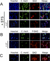
(A) In order to visualize BAD by immunofluorescence, epithelial cells were transfected with the BAD expression plasmid prior to infection. Cells over-expressing BAD were infected with C. trachomatis for 26 h and then incubated with 50 μM LY294002 (LY) or control buffer for 6 h before treating with 2 μM STS or control buffer overnight. BAD is localized to the surface of Chlamydia vacuoles in infected cells, with or without STS treatment (first and second rows). The co-localization of BAD and Chlamydia vacuoles is lost in STS-treated infected cells that had been pre-incubated with LY294002 (bottom row). (B) Cells over-expressing BAD were infected with C. trachomatis for 15 h and then incubated with control buffer (top row) or 50 μM LY294002 (LY) (bottom row) for 6 h. Phosphorylated BAD is concentrated around the early Chlamydia vacuoles, but has a cytosolic distribution in infected cells after treatment with LY294002. Cells treated with LY294002 were exposed at a longer time in order to visualize background phosphorylated-BAD staining. (C) Cells over-expressing BAD were infected with C. muridarum for 15 h. BAD has a cytosolic distribution in infected cells, even in the absence of LY294002. DNA was revealed with Hoechst (blue), Chlamydia with FITC-conjugated anti-Chlamydia antibody (green), and BAD with Texas Red-conjugated anti-BAD antibody or phosphorylated BAD with Texas Red-conjugated antibody recognizing BAD phosphorylated on residue Ser136 (red). In each case, one experiment of two representative experiments performed on separate days is shown.
Figure 8
Figure 8. Localization of 14-3-3β in C. trachomatis-Infected Cells
Cells expressing endogenous levels of BAD were infected with C. trachomatis at an MOI of 1.0 for 26 h, and then incubated with 50 μM LY294002 (LY) or control buffer for 6 h before treating with 2 μM STS overnight. DNA was revealed with Hoechst (blue), Chlamydia with FITC-conjugated anti-Chlamydia antibody (green), and 14-3-3 with Texas Red-conjugated anti-14-3-3 antibody (red), as described in Materials and Methods. 14-3-3 is localized primarily on the Chlamydia vacuole in infected cells, even in the presence of STS (top row). The co-localization of 14-3-3 and the Chlamydia vacuole is lost in infected cells that had been pre-incubated with LY294002 before STS treatment (bottom row). One experiment of three representative experiments performed on separate days is shown.
Figure 9
Figure 9. Summary of the Effects of C. trachomatis Infection on BAD Phosphorylation, BAD Degradation, and Host-Cell Death
Infection with C. trachomatis leads to activation of PI3K, which in turns results in AKT activation and subsequent BAD phosphorylation. In parallel, infection causes degradation of most unphosphorylated BAD. Simultaneous expression of IncG on the chlamydial inclusion and binding of 14-3-3β by IncG allows the inclusion to recruit phosphorylated BAD, preventing the ability of the remaining BAD to translocate to the mitochondria and induce host-cell death.

"VSports app下载" References

    1. Gerbase AC, Rowley JT, Mertens TE. Global epidemiology of sexually transmitted diseases. Lancet. 1998;351:2–4. - PubMed (VSports注册入口)
    1. Miller WC, Ford CA, Morris M, Handcock MS, Schmitz JL, et al. Prevalence of chlamydial and gonococcal infections among young adults in the United States. JAMA. 2004;291:2229–2236. - V体育官网 - PubMed
    1. Thylefors B, Negrel AD, Pararajasegaram R, Dadzie KY. Global data on blindness. Bull World Health Organ. 1995;73:115–121. - PMC - PubMed
    1. Schachter J. Infection and disease epidemiology. In: Stephens RS, editor. Chlamydia: Intracellular biology, pathogenesis, and immunity. Washington (D.C.): ASM Press; 1999. pp. 139–169.
    1. Belland R, Ojcius DM, Byrne GI. Chlamydia . Nature Rev Microbiol. 2004;2:530–531. - PubMed

Publication types

MeSH terms