Skip to main page content
U.S. flag

An official website of the United States government

Dot gov

The . gov means it’s official. Federal government websites often end in . gov or . mil VSports app下载. Before sharing sensitive information, make sure you’re on a federal government site. .

Https

The site is secure V体育官网. The https:// ensures that you are connecting to the official website and that any information you provide is encrypted and transmitted securely. .

Comparative Study
. 2006 May 16:7:115.
doi: 10.1186/1471-2164-7-115.

Cell-type specific gene expression profiles of leukocytes in human peripheral blood

Affiliations
Comparative Study

Cell-type specific gene expression profiles of leukocytes in human peripheral blood (VSports注册入口)

Chana Palmer et al. BMC Genomics. .

"V体育官网入口" Abstract

Background: Blood is a complex tissue comprising numerous cell types with distinct functions and corresponding gene expression profiles VSports手机版. We attempted to define the cell type specific gene expression patterns for the major constituent cells of blood, including B-cells, CD4+ T-cells, CD8+ T-cells, lymphocytes and granulocytes. We did this by comparing the global gene expression profiles of purified B-cells, CD4+ T-cells, CD8+ T-cells, granulocytes, and lymphocytes using cDNA microarrays. .

Results: Unsupervised clustering analysis showed that similar cell populations from different donors share common gene expression profiles. Supervised analyses identified gene expression signatures for B-cells (427 genes), T-cells (222 genes), CD8+ T-cells (23 genes), granulocytes (411 genes), and lymphocytes (67 genes). No statistically significant gene expression signature was identified for CD4+ cells. Genes encoding cell surface proteins were disproportionately represented among the genes that distinguished among the lymphocyte subpopulations. Lymphocytes were distinguishable from granulocytes based on their higher levels of expression of genes encoding ribosomal proteins, while granulocytes exhibited characteristic expression of various cell surface and inflammatory proteins. V体育安卓版.

Conclusion: The genes comprising the cell-type specific signatures encompassed many of the genes already known to be involved in cell-type specific processes, and provided clues that may prove useful in discovering the functions of many still unannotated genes V体育ios版. The most prominent feature of the cell type signature genes was the enrichment of genes encoding cell surface proteins, perhaps reflecting the importance of specialized systems for sensing the environment to the physiology of resting leukocytes. .


Figures
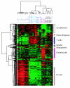
Figure 1
Figure 1
Overview of hierarchical clustering of all samples. Hierarchically clustered gene expression profiles of 28 mixed and purified leukocyte samples. Results for ~741 genes with high variation in transcript levels among these samples. Genes and blood samples are organized by hierarchical clustering based on overall similarity in expression patterns. Expression levels are represented by a color key in which bright red represents the highest levels and bright green represents the lowest levels, and less saturated shades represent intermediate levels of expression. Values were centered across all samples to a mean of zero. Clusters of genes are labeled according to which samples have the highest relative gene expression.
Figure 2
Figure 2
B-cell signature genes. Relative expression of B-cell signature genes is shown for all samples. Genes (rows) are sorted in descending order of correlation of gene expression with relative abundance of B-cells. Samples (columns) are ordered in decreasing order of estimated relative abundance of B-cells from left to right. The grayscale bar on top shows the estimated relative abundance of B-cells in each class of samples – black indicates 100% relative abundance of B-cells, white indicates 0% relative abundance of B-cells, and grey indicates intermediate relative abundance of B-cells. Gene expression values are centered across all samples to a median of zero. All genes mentioned in the text are listed in order of correlation with B-cell abundance and black bars indicate their positions in the figure.
Figure 3
Figure 3
T-cell signature genes. Relative expression of T-cell signature genes is shown for all samples. Genes (rows) are sorted in descending order of correlation of gene expression with relative abundance of T-cells. Samples (columns) are ordered in decreasing relative abundance from left to right. Gene expression values are centered across all samples to a median of zero. The grayscale bar on top shows the estimated relative abundance of T-cells in each class of samples – black indicates 100% relative abundance of T-cells, white indicates 0% relative abundance of T-cells, and grey indicates intermediate relative abundance of T-cells. All genes mentioned in the text are listed in order of correlation with T-cell abundance, and black bars indicate their positions in the figure.
Figure 4
Figure 4
CD8+ T-cell signature genes. Relative expression of CD8+ T-cell signature genes is shown for all samples. Genes (rows) are sorted in descending order of correlation of gene expression with relative abundance of T-cells. Samples (columns) are ordered in decreasing relative abundance from left to right. The grayscale bar on top shows the estimated relative abundance of CD8+ T-cells in each class of samples – black indicates 100% relative abundance of CD8+ T-cells, white indicates 0% relative abundance of CD8+ T-cells, and grey indicates intermediate relative abundance of CD8+ T-cells. Gene expression values are centered across all samples to a median of zero. All genes mentioned in the text are listed and their position is marked with a black bar.
Figure 5
Figure 5
Granulocyte signature genes. Relative expression of granulocyte signature genes is shown for all samples. Genes (rows) are sorted in descending order of correlation of gene expression with relative abundance of granulocytes. Samples (columns) are ordered in decreasing relative abundance from left to right. The grayscale bar on top shows the estimated relative abundance of granulocytes in each class of samples – black indicates 100% relative abundance of granulocytes, white indicates 0% relative abundance of granulocytes, and grey indicates intermediate relative abundance of granulocytes. Gene expression values are centered across all samples to a median of zero. All genes mentioned in the text are listed in order of correlation with granulocyte abundance, and black bars indicate their positions in the figure.
Figure 6
Figure 6
Lymphocyte signature genes. Relative expression of lymphocyte signature genes is shown for all samples. Genes (rows) are sorted in ascending order of descending correlation of gene expression with relative abundance of lymphocytes. Samples (columns) are ordered in increasing relative abundance from left to right. The grayscale bar on top shows the estimated relative abundance of lymphocytes in each class of samples – black indicates 100% relative abundance of lymphocytes, white indicates 0% relative abundance of lymphocytes, and grey indicates intermediate relative abundance of lymphocytes. Gene expression values are centered across all samples to a median of zero. All genes mentioned in the text are listed and their position is marked with a black bar.

References

    1. Parks D, Herzenberg L. Fluorescence-activated cell sorting: Theory, experimental optimization, and applications in lymphoid cell biology. Methods Enzymol. 1984;108:197–241. - PubMed
    1. Bomprezzi R, Ringner M, Kim S, Bittner ML, Khan J, Chen Y, Elkahloun A, Yu A, Bielekova B, Meltzer PS, Martin R, McFarland HF, Trent JM. Gene expression profile in multiple sclerosis patients and healthy controls: identifying pathways relevant to disease. Hum Mol Genet. 2003;12:2191–2199. doi: 10.1093/hmg/ddg221. - DOI - PubMed
    1. Connolly PH, Caiozzo VJ, Zaldivar F, Nemet D, Larson J, Hung SP, Heck JD, Hatfield GW, Cooper DM. Effects of exercise on gene expression in human peripheral blood mononuclear cells. J Appl Physiol. 2004;97:1461–1469. doi: 10.1152/japplphysiol.00316.2004. - "VSports最新版本" DOI - PubMed
    1. Lampe JW, Stepaniants SB, Mao M, Radich JP, Dai H, Linsley PS, Friend SH, Potter JD. Signatures of environmental exposures using peripheral leukocyte gene expression: tobacco smoke. Cancer Epidemiol Biomarkers Prev. 2004;13:445–453. - VSports在线直播 - PubMed
    1. Moore DF, Li H, Jeffries N, Wright V, Cooper RA, Jr, Elkahloun A, Gelderman MP, Zudaire E, Blevins G, Yu H, Goldin E, Baird AE. Using peripheral blood mononuclear cells to determine a gene expression profile of acute ischemic stroke: a pilot investigation. Circulation. 2005;111:212–221. doi: 10.1161/01.CIR.0000152105.79665.C6. - DOI - PubMed

Publication types

MeSH terms