<b dir="wccbfu8"></b> "V体育ios版" Skip to main page content
U.S. flag

An official website of the United States government

Dot gov

The . gov means it’s official. Federal government websites often end in VSports app下载. gov or . mil. Before sharing sensitive information, make sure you’re on a federal government site. .

Https

The site is secure. The https:// ensures that you are connecting to the official website and that any information you provide is encrypted and transmitted securely V体育官网. .

. 2006 Apr;2(4):e58.
doi: 10.1371/journal.pgen.0020058. Epub 2006 Apr 21.

"VSports app下载" Loss of Atrx affects trophoblast development and the pattern of X-inactivation in extraembryonic tissues

Affiliations

V体育安卓版 - Loss of Atrx affects trophoblast development and the pattern of X-inactivation in extraembryonic tissues

David Garrick et al. PLoS Genet. 2006 Apr.

"V体育ios版" Abstract

ATRX is an X-encoded member of the SNF2 family of ATPase/helicase proteins thought to regulate gene expression by modifying chromatin at target loci. Mutations in ATRX provided the first example of a human genetic disease associated with defects in such proteins. To better understand the role of ATRX in development and the associated abnormalities in the ATR-X (alpha thalassemia mental retardation, X-linked) syndrome, we conditionally inactivated the homolog in mice, Atrx, at the 8- to 16-cell stage of development. The protein, Atrx, was ubiquitously expressed, and male embryos null for Atrx implanted and gastrulated normally but did not survive beyond 9 VSports手机版. 5 days postcoitus due to a defect in formation of the extraembryonic trophoblast, one of the first terminally differentiated lineages in the developing embryo. Carrier female mice that inherit a maternal null allele should be affected, since the paternal X chromosome is normally inactivated in extraembryonic tissues. Surprisingly, however, some carrier females established a normal placenta and appeared to escape the usual pattern of imprinted X-inactivation in these tissues. Together these findings demonstrate an unexpected, specific, and essential role for Atrx in the development of the murine trophoblast and present an example of escape from imprinted X chromosome inactivation. .


Conflict of interest statement

Competing interests. The authors have declared that no competing interests exist V体育安卓版.

VSports最新版本 - Figures

Figure 1
Figure 1. Schematic Representation of the ATRX Isoforms
Shown at the top is the human ATRX cDNA. The boxes represent the 36 exons. The introns are not to scale. The alternative splicing of exons 6 and 7 is indicated. Shown underneath are the two ATRX protein isoforms. Full-length ATRX (~280 kDa) is encoded by the largest open reading frame. The positions of the principal features (the PHD-like domain and the seven SWI/SNF-like motifs) are indicated. Above full-length ATRX is shown the truncated ATRXt isoform (apparent molecular weight of ~200 kDa) that arises through the failure to splice intron 11 and the use of an intronic poly(A) signal. The intron-encoded region of ATRXt is indicated as a filled grey box. Locations of recombinant proteins (A2, FXNP5, and H-300) used to generate antibodies are shown. The scale bar represents 200 amino acids.
Figure 2
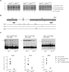
Figure 2. Cre-Mediated Ablation of Full-Length Atrx Protein in ES Cells
(A) Strategy for targeted deletion of exon 18 of the Atrx gene. The top line shows the wild-type allele (Atrx WT) at the region surrounding exon 18. Below is shown the targeting vector and the targeted allele (Atrx flox) resulting from homologous recombination. The loxP target sites of the Cre recombinase are shown as black triangles, and the three possible recombination events that can be mediated by the Cre recombinase are indicated (labelled A, B, and C in the Atrx flox allele). At bottom is shown the Cre-recombined allele (Atrx Δ18Δneo) (resulting from recombination event C) in which both exon 18 and the MC1neopA selection cassette have been deleted. EcoRI (labelled E) and SacI (labelled S) sites present on the targeted 129 strain X chromosome are indicated. Black bars indicate the positions of the probes used in Southern blots. (B) Southern blot analysis of EcoRI-digested DNA from either wild-type ES cells (E14) or targeted ES cell clones bearing the Atrx flox allele (1/F12 and 1/G11) hybridised with either the 20/27 (left blot) or Hae0.9 (right blot) probes. The EcoRI fragment of the Atrx WT allele (18.5 kb) has been replaced with the expected fragments of 11.2 kb (20/27 probe) or 8.5 kb (Hae0.9 probe) (C) Southern blot analysis of SacI-digested DNA from either wild-type ES cells (E14) or targeted ES cell clones bearing the Atrx flox allele (1/F12 and 1/G11) or Cre-recombinant clones derived from these (1/F12B1F12 and 1/G11D5). The membrane was hybridised with the intron 17 probe indicated in (A). The expected bands of 6.2 (Atrx WT), 5.0 (Atrx flox), and 2.8 (Atrx Δ18Δneo) kb were observed. (D) Northern blot analysis of RNA from ES cells shown in (C). The membrane was hybridised first to a probe from exon 10 of the Atrx gene (top blot) and subsequently to a β-actin cDNA probe as loading control (bottom blot). The transcripts responsible for full-length Atrx (~10 kb) and the truncated Atrxt isoforms (~7 kb) are indicated. (E) Western blot analysis of whole-cell extracts from the clones shown in (C) using an anti-ATRX monoclonal antibody (23C, raised against peptide A2 of the human ATRX protein shown in Figure 1). The full-length and truncated Atrx isoforms are indicated.
Figure 3
Figure 3. Growth and Methylation Defects in Atrxnull ES Cells
(A) Cultures were inoculated with equivalent numbers of ES cells bearing different Atrx alleles as indicated, and were serially passaged. After the indicated days of coculture, DNA extracted from a sample of cells was analysed by Southern blot to detect the Atrx alleles. DNA was digested with SpeI, and the membrane was hybridised with the 20/27 probe shown in Figure 2A. The expected sizes of the different alleles are indicated. (B) Schematic diagram of the transcribed portion of the mouse rDNA repeat with the 18S, 5.8S, and 28S genes indicated. The positions of the limit-digesting enzymes BamH (labelled B) and EcoRI (labelled E) and the probes (RIB3 and RIB4) used in the Southern blots shown in (C) are indicated. Below are shown the locations of the methylation-sensitive enzymes (SmaI, PvuI, and MluI) whose methylation status has been analysed in the Southern blots shown in (C). (C) DNA from Atrx-positive (Atrx+, bearing either an Atrx WT or Atrx flox allele) or Atrxnull (bearing the Atrx Δ18Δneo allele) ES cells and 7-d embryoid bodies were digested with the enzymes shown and analysed by Southern blotting using the probes indicated. Arrows indicate the fully methylated copies (cut by only the limit-digesting enzyme). Phosphorimager quantitation of the blots are shown below. The y-axis shows the percentage of copies that are undigested by the methylation-sensitive enzyme as a percentage of the total signal from cut and uncut rDNA. Mean values are indicated by horizontal lines, and the significance of the differences between the Atrx-positive and Atrxnull populations are shown for each enzyme.
Figure 4
Figure 4. Timing of Onset of GATA1-Cre Expression and PCR Genotyping of Atrx Alleles
(A) GATA1-cre +/+ transgenic males were crossed to females of the ROSA26 reporter strain (ROSA26 +/), and embryos were recovered at 0.5 dpc (~16-cell morula stage) and stained with X-gal. Cre-mediated activation of the ROSA26 β-galactosidase reporter allele was detected in all cells in embryos in which both alleles are coinherited. (B) Top gel: PCR genotyping of Atrx alleles in embryos using primers PPS1.15 (exon 17) and Mxnp30 (exon 20) as described in Protocol S1. The sizes of PCR products from the different alleles are indicated. Both the Atrx Δ18 (resulting from recombination event B in Figure 2A) and the Atrx Δ18Δneo allele (resulting from recombination event C in Figure 2A) are null for full-length Atrx protein. The bottom gel shows products from a PCR reaction (primers DG52/DG53) used to sex embryos as described in Protocol S1. A 450-bp PCR product is amplified from a mouse Y chromosome-specific satellite repeat.
Figure 5
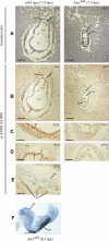
Figure 5. Morphology of Atrxnull Embryos at 7.5 dpc and 8.5 dpc
Paraffin sections of wild-type or Atrxnull 7.5 dpc embryos (dissected in their deciduas) were stained with haematoxylin (A) or with an anti-ATRX antibody (H-300, Figure 1) (B–E). Photomicrographs C–E show higher magnification images (200×) of the stained sections shown in (B) (40×). Scale bars represent 200 μm (40× magnification) or 40 μm (200× magnification). a, amnion; ac, amniotic cavity; c, chorion; e, epiblast; ec, ectoplacental cavity; ecc, exocoelomic cavity; ep, ectoplacental cone; ne, neural ectoderm; rm, Reichert's membrane; tgc, trophoblast giant cell. (F) Detection of brachyury (T) expression in Atrxnull 8.5 dpc embryo (head fold stage) by WMISH. The genotype was determined by PCR (as shown in Protocol S1) using DNA extracted from yolk sac. hf, head fold; n, emerging notochord; ps, primitive streak.
Figure 6
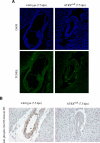
Figure 6. Analysis of Apoptosis and Mitosis in Atrxnull Embryos
(A) Paraffin sections of wild-type or Atrxnull 7.5 dpc embryos (dissected in their deciduas) were analysed by TUNEL assay and apoptotic cells labelled with fluorescein-dUTP. Sections were counterstained with DAPI. (B) Paraffin sections of wild-type or Atrxnull 7.5 dpc embryos were stained with an antibody against the mitosis marker phosphorylated (Ser10) histone H3. Sections were counterstained with haematoxylin. For both (A) and (B), the presence or absence of Atrx in each embryo was determined by staining adjacent sections with the anti-ATRX antibody (H-300) as in Figure 5 (unpublished data).
Figure 7
Figure 7. Trophectoderm Defect in Atrxnull Embryos
(A) 8.5 dpc embryos were dissected from surrounding decidual tissue and observed in whole mount. The genotype of each (indicated above) was determined by PCR using DNA extracted from whole embryos after photography. In the left image, the wild-type female (three-somite stage, left) is surrounded by trophoblast (t) while the trophoblast component surrounding the Atrxnull males (at headfold/presomite [middle] and two-somite stages [right], respectively) is severely depleted. In the right image, the trophoblast has been dissected away from the embryonic region of the wild-type embryo, to reveal the small, abnormally shaped ectoplacental cone (epc) of the mutant littermates. (B) WMISH to detect expression of Pl-1 (a marker of TGCs) at the implantation sites in vacated deciduas that had contained 8.5 dpc wild-type (Atrx WT/WT) or Atrxnull (Atrx Δ18Δneo/Y) embryos. The genotype was determined by PCR using DNA extracted from whole embryos. TGCs are stained with Pl-1. (C) Paraffin sections of wild-type or Atrxnull 7.5 dpc embryos (dissected in their deciduas) were stained with an anti-Pl-1 antibody. The presence or absence of Atrx in each embryo was determined by staining adjacent sections with the anti-ATRX antibody (H-300) as in Figure 5 (unpublished data). (D) Examples of 5-d blastocyst outgrowth cultures. Extensive trophoblast outgrowing from the inner cell mass (icm) was observed in all genotypes. The Atrx genotype and sex of the blastocyst indicated were determined by PCR.
Figure 8
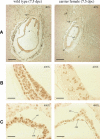
Figure 8. Escape from Imprinted Inactivation of the Paternally Inherited AtrxWT Allele in Carrier Females
Paraffin sections of wild-type (Atrx WT/Y) and carrier female (Atrx WT/null) 7.5 dpc embryos (dissected in their deciduas) were stained with the anti-ATRX antibody (H-300). Scale bars represent 200 μm (40× magnification) or 20 μm (400× magnification). (A) Stained sections showing whole embryos at 40× magnification. a, amnion; c, chorion; e, epiblast; ep, ectoplacental cone. (B) Higher-magnification image (400×) of the epiblast regions of the stained sections shown in (A). (C) Higher-magnification image (400×) showing the extraembryonic derived-chorionic ectoderm of the stained sections shown in (A). ce, chorionic ectoderm; cm, chorionic mesoderm.

References

    1. Gibbons RJ, Higgs DR. Molecular-clinical spectrum of the ATR-X syndrome. Am J Med Genet. 2000;97:204–212. - PubMed
    1. Gibbons RJ, Suthers GK, Wilkie AO, Buckle VJ, Higgs DR. X-linked alpha-thalassemia/mental retardation (ATR-X) syndrome: Localization to Xq12-q21.31 by X inactivation and linkage analysis. Am J Hum Genet. 1992;51:1136–1149. - V体育ios版 - PMC - PubMed
    1. Gibbons RJ, Picketts DJ, Villard L, Higgs DR. Mutations in a putative global transcriptional regulator cause X-linked mental retardation with alpha-thalassemia (ATR-X syndrome) Cell. 1995;80:837–845. - PubMed
    1. Garrick D, Samara V, McDowell TL, Smith AJ, Dobbie L, et al. A conserved truncated isoform of the ATR-X syndrome protein lacking the SWI/SNF-homology domain. Gene. 2004;326:23–34. - PubMed
    1. Picketts DJ, Tastan AO, Higgs DR, Gibbons RJ. Comparison of the human and murine ATRX gene identifies highly conserved, functionally important domains. Mamm Genome. 1998;9:400–403. - PubMed

Publication types

MeSH terms