Skip to main page content
U.S. flag

An official website of the United States government

Dot gov

The . gov means it’s official. Federal government websites often end in . gov or . mil VSports app下载. Before sharing sensitive information, make sure you’re on a federal government site. .

Https

The site is secure. The https:// ensures that you are connecting to the official website and that any information you provide is encrypted and transmitted securely V体育官网. .

. 2006 Apr;116(4):929-39.
doi: 10.1172/JCI27363. Epub 2006 Mar 23.

"V体育官网" GATA-6 regulates semaphorin 3C and is required in cardiac neural crest for cardiovascular morphogenesis

Affiliations

GATA-6 regulates semaphorin 3C and is required in cardiac neural crest for cardiovascular morphogenesis

John J Lepore et al. J Clin Invest. 2006 Apr.

Abstract

GATA transcription factors play critical roles in restricting cell lineage differentiation during development. Here, we show that conditional inactivation of GATA-6 in VSMCs results in perinatal mortality from a spectrum of cardiovascular defects, including interrupted aortic arch and persistent truncus arteriosus. Inactivation of GATA-6 in neural crest recapitulates these abnormalities, demonstrating a cell-autonomous requirement for GATA-6 in neural crest-derived SMCs. Surprisingly, the observed defects do not result from impaired SMC differentiation but rather are associated with severely attenuated expression of semaphorin 3C, a signaling molecule critical for both neuronal and vascular patterning. Thus, the primary function of GATA-6 during cardiovascular development is to regulate morphogenetic patterning of the cardiac outflow tract and aortic arch. These findings provide new insights into the conserved functions of the GATA-4, -5, and -6 subfamily members and identify GATA-6 and GATA-6-regulated genes as candidates involved in the pathogenesis of congenital heart disease VSports手机版. .


Figures

Figure 1
Figure 1. Conditional targeting of murine GATA-6.
(A, top panel) GATA-6 locus containing exons 3 and 4 (rectangles) and the targeting construct containing phosphoglycerate kinase–regulated (PGK-regulated) neo and HSV thymidine kinase (tk) genes. loxP sites (triangles) flank neo and exon 4 encoding the C-terminal zinc finger (Zn) DNA-binding domain. B, BamHI. (Middle panel) Conditionally targeted GATA-6 allele. (Bottom panel) Targeted allele following selective neo deletion. (B, left) Southern blotting (probe A) of targeted ES cells identifies wild-type (11.1 kb) and conditionally targeted (6.8 kb) alleles. (Right) Southern blotting (probe B) of ES cells following Cre transfection identifies wild-type (11.1 kb) and conditionally targeted alleles with neo (6.2 kb) and with selective neo deletion (4.3 kb). (C) Genotyping of wild-type (+/+), heterozygous (+/F), and homozygous (F/F) conditionally targeted mice. (Left) Southern blotting (probe B) identifies wild-type (11.1 kb) and conditionally targeted (4.3 kb) alleles. (Right) PCR using primers PCR-A and PCR-B identifies products corresponding to wild-type (150 bp) and targeted (110 bp) alleles. (D) Analysis of primary GATA-6F/F aortic SMCs infected with Ad-empty or Ad-Cre. RT-PCR identifies products corresponding to wild-type GATA-6 (373 bp) and GATA-6 following exon 4 deletion (285 bp). Western blotting (WB) identifies full-length (45 kDa) and truncated (41 kDa) GATA-6 proteins. (E) Activation of the GATA-dependent Dab2-LUC reporter. NIH3T3 cells were transiently transfected with 100 ng of the Dab2-LUC reporter and with 0.1–2.0 μg of either pcDNA3–GATA-6 or pcDNA3–GATA-6–Δexon4. The reporter was activated by expression of increasing amounts of wild-type GATA-6, but not by expression of the truncatedATA-6–Δexon4 protein.
Figure 2
Figure 2. Cardiovascular abnormalities produced by conditional GATA-6 deletion.
Aortic arch patterning and cardiac outflow tract septation were examined in E18.5 GATA-6F/F(Cre–; A and FI), SMCre+GATA-6F/F (SMCre+; B, C, and KN), and Wnt1Cre+GATA-6 (WCre+; D, E, J, and O) embryos using polymer vascular casting (AE) and H&E staining (FO). (A) In normal aortic arch patterning, the ascending aorta (AAo) and pulmonary artery (PA) are distinct, septated vessels. The ductus arteriosus (DA) is patent and connects the PA to the proximal descending aorta (DAo). The right subclavian artery (RS) branches from the AAo. (B) SMCre+ embryo demonstrating truncus arteriosus (TA) and hypoplastic aortic arch (arch). (C) SMCre+ embryo demonstrating interrupted aortic arch (IAA). (D) WCre+ embryo demonstrating TA, hypoplastic arch, and retroesophageal right subclavian artery (RERS). (E) WCre+ embryo demonstrating IAA. (FI) Serial histological sections through the heart and great vessels of a normal embryo. (F) The DA connects the PA to the DAo. The RS branches from the AAo. (G and H) The AAo and PA are distinct, septated vessels. The pulmonary valve (PV) is shown. (I) An intact ventricular septum (arrow) separates the right and left ventricles. (KN) Corresponding serial sections from a SMCre+ embryo. (K) A RERS branches from the DAo and travels posterior to the esophagus (E). (LN) There is single outflow tract vessel, or TA; a single, common aorticopulmonary valve (APV); and a membranous ventricular septal defect (VSD). (J and O) Representative WntCre+ embryo exhibiting TA and VSD. Original magnification, ×20 (AE); ×40 (FO).
Figure 3
Figure 3. Expression of GATA-6 in cardiac neural crest derivatives.
GATA-6 expression was examined by in situ hybridization in wild-type, SM22CreGATA-6F/F (SMCre), and SM22Cre+GATA-6F/F (SMCre+) embryos. (A and B) In wild-type embryos at E9.5, GATA-6 is expressed in the cardiac outflow tract (OFT), atria (At), bulbus cordis (BC), and left ventricle. (C and D) In wild-type embryos at E11.5, GATA-6 is expressed in the SMCs (C, arrows) that populate the vascular wall of the AAo and PA, in the conotruncal endocardial cushions in the regions populated by migrating neural crest cells (D, long arrows) and in the cuff of cardiac myocytes at the base of the outflow tract (D, short arrow). (E and F) In situ hybridization studies with a GATA-6 exon 4 probe demonstrate GATA-6 expression in the OFT, At, and right and left ventricles that is markedly attenuated in E11.5 SMCre+ embryos (F) as compared with SMCre embryos (E). (G) In wild-type embryos at E12.5, GATA-6 is abundantly expressed in neural crest–derived SMCs populating the aorta (Ao), PA, and DA; in cardiac myocytes in the At, right ventricle, and left ventricle; and in the endocardial cushions (EC). Original magnification, ×100 (A, B, and EG); ×200 (C and D).
Figure 4
Figure 4. Effect of GATA-6 deletion on neural crest–derived SMC differentiation.
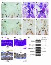
(AJ) H&E staining (C, D, G, and H) and SMα-actin (SMA) immunohistochemistry (A, B, E, F, I, and J) of SM22CreGATA-6F/F (SMCre) and SM22Cre+GATA-6F/F (SMCre+) embryos. Original magnification, ×100 (AF); ×200 (GJ). (A and B) Frontal section through the pharyngeal arches in an E11.5 SMCre+ embryo (B) demonstrates normal formation of pharyngeal arches 3, 4, and 6 and SMA immunostaining in SMCs of the pharyngeal arch arteries and carotid arteries (car) similar to that in a normal SM22Cre embryo (A). (CF) In a representative E12.5 SMCre+ embryo (D and F), despite the presence of aortopulmonary window (APW), the structure of the vessel wall and the presence of SMA immunostaining in neural crest–derived SMCs in the vascular wall of the aorta and PA are not different compared with a normal SMCre embryo (C and E). (GJ) Cross sections through the vascular walls of Ao and PA of an E18.5 SM22Cre embryo (G and I) and the vascular wall of the truncus arteriosus (TA) of a SM22Cre+ embryo (H and J) demonstrate similar structure of the vessel wall and presence of SMA immunostaining neural crest–derived SMCs. (K) Quantitative RT-PCR of primary GATA-6F/F aortic SMCs infected with Ad-empty (Ctrl) or Ad-Cre (Cre) demonstrates that GATA-6 deletion does not change expression of mRNA encoding SMC-restricted cytoskeletal and contractile proteins. The ethidium-stained products amplified at the cycle threshold for each message and for control GAPDH are shown with the fold change in expression.
Figure 5
Figure 5. Transcriptional regulation of semaphorin 3C (sema 3C) by GATA-6.
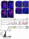
(AH) Semaphorin 3C and plexin A2 in situ hybridization. In E12.5 SM22CreGATA-6F/F (SMCre) embryos, semaphorin 3C (A) and plexin A2 (C) are abundantly expressed in neural crest–derived SMCs populating the aorta, PA, DA, and cuff of surrounding myocardial cells (arrows). In SMCre+ embryos, SMC expression of semaphorin 3C (B) is markedly reduced whereas plexin A2 expression (D) is not. Semaphorin 3C– (E) and plexin A2– (G) expressing neural crest cells are identified within the conotruncal endocardial cushions (arrows) in E11.5 Wnt1CreGATA-6F/F (WntCre) embryos, but semaphorin 3C and plexin A2 expression is not observed in WCre+ embryos (F and H, respectively). Original magnification, ×100 (AD); ×200 (EH). (I) VISTA comparison of murine and human semaphorin 3C proximal promoter, exon 1 (ex 1), and exon 2. The x and y axes indicate sequence length (kb) and percentage of homology (≥ 75%, pink), respectively. GATA-binding sites conserved in mouse, rat, and human sequence are indicated by asterisks. (J) Schematic of the sema3C-LUC reporter construct containing the 0.9-kb proximal promoter, exon 1, intron 1, and 35 bp of exon 2 upstream of firefly luciferase (LUC). (K) Activation of the sema3C-LUC reporter by GATA-6. NIH3T3 cells were transiently transfected with 100 ng of sema3C-LUC and with 1–5 μg of expression plasmid encoding wild-type GATA-6, GATA-6 containing zinc finger mutations abrogating DNA binding (GATA-6 mut), or GATA-6 lacking sequences encoded by exon 4 (GATA-6 Δexon4). The reporter was activated by expression of wild-type GATA-6 but not by expression of GATA-mut or GATA-6 Δexon4.

"VSports" References

    1. Gruber P.J., Epstein J.A. Development gone awry: congenital heart disease. Circ. Res. 2004;94:273–283. - PubMed (VSports最新版本)
    1. Creazzo T.L., Godt R.E., Leatherbury L., Conway S.J., Kirby M.L. Role of cardiac neural crest cells in cardiovascular development. Annu. Rev. Physiol. 1998;60:267–286. - PubMed
    1. Stoller J.Z., Epstein J.A. Cardiac neural crest. Semin. Cell Dev. Biol. . 2005;16:704–715. - PubMed
    1. Feiner L., et al. Targeted disruption of semaphorin 3C leads to persistent truncus arteriosus and aortic arch interruption. Development. 2001;128:3061–3070. - PubMed
    1. Brown C.B., et al. PlexinA2 and semaphorin signaling during cardiac neural crest development. Development. 2001;128:3071–3080. - "V体育官网入口" PubMed

Publication types (VSports)

V体育安卓版 - MeSH terms