Skip to main page content
U.S. flag

An official website of the United States government

Dot gov

The . gov means it’s official. Federal government websites often end in . gov or . mil VSports app下载. Before sharing sensitive information, make sure you’re on a federal government site. .

Https

The site is secure. The https:// ensures that you are connecting to the official website and that any information you provide is encrypted and transmitted securely. V体育官网.

. 2006 Feb 14;103(7):2380-5.
doi: 10.1073/pnas.0506668103. Epub 2006 Feb 7.

Streptococcal modulation of cellular invasion via TGF-beta1 signaling

Affiliations

Streptococcal modulation of cellular invasion via TGF-beta1 signaling

Beinan Wang (VSports注册入口) et al. Proc Natl Acad Sci U S A. .

Abstract

Group A Streptococcus (GAS) and other bacterial pathogens are known to interact with integrins as an initial step in a complex pathway of bacterial ingestion by host cells. Efficient GAS invasion depends on the interaction of bound fibronectin (Fn) with integrins and activation of integrin signaling. TGF-beta1 regulates expression of integrins, Fn, and other extracellular matrix proteins, and positively controls the integrin signaling pathway. Therefore, we postulated that TGF-beta1 levels could influence streptococcal invasion of mammalian cells. Pretreatment of HEp-2 cells with TGF-beta1 increased their capacity to ingest GAS when the bacteria expressed fibronectin-binding proteins (M1 or PrtF1). Western blots revealed significant induction of alpha5 integrin and Fn expression by HEp-2 cells in response to TGF-beta1 VSports手机版. Increased ingestion of streptococci by these cells was blocked by a specific inhibitor of the TGF-beta1 receptor I and antibodies directed against alpha5 integrin and Fn, indicating that increased invasion depends on TGF-beta1 up-regulation of both the alpha5 integrin and Fn. The capacity of TGF-beta1 to up-regulate integrin expression and intracellular invasion by GAS was reproduced in primary human tonsil fibroblasts, which could be a source of TGF-beta1 in chronically infected tonsils. The relationship between TGF-beta1 and GAS invasion was strengthened by the observation that TGF-beta1 production was stimulated in GAS-infected primary human tonsil fibroblasts. These findings suggest a mechanism by which GAS induce a cascade of changes in mammalian tissue leading to elevated expression of the alpha5beta1 receptor, enhanced invasion, and increased opportunity for survival and persistence in their human host. .


Conflict of interest statement

Conflict of interest statement: No conflicts declared.

Figures

Fig. 1.
Fig. 1.
TGF-β1 enhances GAS invasion of epithelial cells. HEp-2 cells were pretreated with TGF-β1 and infected with strains 90-226 (M1+PrtF1) and JRS4 (M6+ PrtF1+). Extracellular bacteria were killed by 2-h incubation with antibiotics. Intracellular bacteria were released by lysis buffer and quantified by colony-forming units (CFU). CFU from nontreated control cells (Ctrl) was considered as 100%. CFU from treated cells was normalized against controls and was reported as a percent of the control. Numbers shown above the columns are percentages of inoculated CFU internalized. Data are presented as means ± SD (n = 3) from a representative of at least three separate experiments. ∗, P < 0.008; ∗∗, P < 0.004 compared with Ctrl.
Fig. 2.
Fig. 2.
TGF-β1 enhanced invasion depends on expression of either M1 or PrtF1 Fn-binding proteins. HEp-2 cells were pretreated with TGF-β1. Invasion assays were performed with 90-226 strains: 90-226 (M1+PrtF1), M1 deletion mutant (M1PrtF1), and PrtF1+ recombinant (M1PrtF1+). Data and numbers above the columns are presented as described in Fig. 1 ∗, P < 0.03; ∗∗, P < 0.05 compared with Ctrl.
Fig. 3.
Fig. 3.
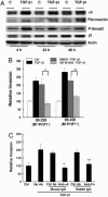
TGF-β1 up-regulates expression of α5 integrin and Fn by epithelial cells. (A) HEp-2 cells were pretreated with TGF-β1 and lysed in RIPA buffer (Supporting Text, which is published as supporting information on the PNAS web site) at indicated time. Equal amounts of protein were separated by SDS∕PAGE and blotted with anti-α5, anti-Fn, anti-phospho-smad2 (P-Smad 2), anti-β1, or anti-actin. (B) HEp-2 cells were not treated (Ctrl) or treated with TGF-β1 for 24 h after pretreatment with the inhibitor of TGF-β1 type I receptor SB 431542 (10 μM), (SB 431542∕TGF-β1) or DMS (10 μM) (inhibitor solvent control), (DMSO∕TGF-β1) or medium alone (TGF-β1) for 60 min, then infected with streptococci. Data are presented as described in Fig. 1. ∗, P < 0.02; ∗∗, P < 0.04. (C) HEp-2 cells were not treated (Ctrl) or pretreated with TGF-β1 and then incubated with anti-α5 integrin and anti-Fn and indicated corresponding control antibodies for 30 min before infection with bacteria. ∗, P < 0.02; ∗∗, P < 0.04 compared with Ctrl.
Fig. 4.
Fig. 4.
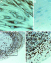
Immunocytochemical detection of vimentin-positive cells. Monolayers of PHTF and 5-μm sections of normal human palatine tonsil were processed for routine immunocytochemistry using a monoclonal antibody against vimentin and a peroxidase-conjugated secondary antibody as described (30). A positive signal is indicated by the brown stain; cell nuclei were counterstained with hematoxylin. (Original magnification: ×400 in A, B, and D and ×100 in C.) (A) PHTF monolayer stained with antivimentin antibody. (B) PHTF monolayer stained with pan-cytokeratin specific antibody. (C) Tonsil section stained with anti-vimentin antibody. (D) Enlargement of area enclosed within the box in C.
Fig. 5.
Fig. 5.
TGF-β1 enhances expression of α5 integrin and invasion of PHTF. (A) PHTF were treated with TGF-β1 and analyzed as described in Fig. 3A. The bands of α5, Fn, and actin were quantified with densitometry. (B and C) Each column shows the ratio of α5 to actin (B) or the ratio of Fn to actin (C). D, days of cells incubated with TGF-β1 before preparation of cell lysate.
Fig. 6.
Fig. 6.
TGF-β1 enhances invasion of PHTF. (A) PHTF were pretreated with TGF-β1. Invasion assays were performed with strain JRS4, (M6+ PrtF1+). Ctrl indicates invasion of cells that were not treated with TGF-β1. Data were normalized and presented as described in Fig. 1. ∗, P < 0.02. (B) PHTF were pretreated with TGF-β1. Cells were infected with strain 90-226 (M1+ PrtF1) in media containing the indicated concentrations of Fn. Percentage invasion equals internalized CFU divided by total CFU inoculated into wells, multiplied by 100. ∗, P < 0.003 compared with Ctrl.
Fig. 7.
Fig. 7.
GAS-induced production of TGF-β1 by PHTF. (A) Cell-free supernatants from PHTF, uninfected, and infected cells with viable strain 90-226 (M1+PrtF1) at a MOI of 1:5 were collected at days 2, 7, and 14 (2D, 7D, and 14D) after infection and heat-activated (90°C, 5 min). Total TGF-β1 activity (active and latent forms) was measured by incubation of supernatants with Mv1Lu reporter cells overnight at 37°C. Cells were then washed and lysed, and luciferase activity in lysates was measured. ∗, P < 0.007; ∗∗, P < 0.004 compared with Ctrl. (B) Supernatants were preincubated with mouse anti-TGF-β1 antibody or nonimmune antibody of the same isotype before addition to Mv1Lu reporter cells. Data are presented as average luciferase activity of triplicate samples ± SD. ∗, P < 0.0001 compared with Ctrl IgG.
Fig. 8.
Fig. 8.
TGF-β1 activation of group A streptococcal ingestion in the tonsils. Interaction of streptococci (chains of banded spheres) with integrin-bound fibronectin or other cellular molecules leads to induction of TGF-β1 from fibroblasts. TGF-β1 then up-regulates expression of α5 integrin (gray tabs) and Fn (open tabs) on epithelial cells and also fibroblasts with formation of a higher density of α5β1 receptors and more efficient ingestion of streptococci. Hatched tab, β1 integrin; solid line, interstitium; dashed arrow, expression of TGF-β1; solid arrow, effect of TGF-β1.

References

    1. Kaplan E. L., Johnson D. R. Pediatrics. 2001;108:1180–1186. - PubMed
    1. Gerber M. A., Tanz R. R., Kabat W., Bell G. L., Siddiqui B., Lerer T. J., Lepow M. L., Kaplan E. L., Shulman S. T. Pediatrics. 1999;104:911–917. - PubMed
    1. Osterlund A., Popa R., Nikkila T., Scheynius A., Engstrand L. Laryngoscope. 1997;107:640–647. - PubMed
    1. Park H. S., Francis K. P., Yu J., Cleary P. P. J. Immunol. 2003;171:2532–2537. - PubMed
    1. Bisno A. L., Brito M. O., Collins C. M. Lancet Infect. Dis. 2003;3:191–200. - V体育官网入口 - PubMed

Publication types

MeSH terms

Substances (V体育官网)

LinkOut - more resources