<u date-time="yLPlZ64"></u><sub date-time="Ep5p2SRO"></sub><sub date-time="slAybw"><small dropzone="LR2ZYK7M"></small></sub> Skip to main page content (V体育2025版)
U.S. flag

An official website of the United States government

Dot gov

The . gov means it’s official. Federal government websites often end in . gov or VSports app下载. mil. Before sharing sensitive information, make sure you’re on a federal government site. .

Https

The site is secure V体育官网. The https:// ensures that you are connecting to the official website and that any information you provide is encrypted and transmitted securely. .

Comparative Study
. 2005 Dec;25(24):10731-44.
doi: 10.1128/MCB.25.24.10731-10744.2005.

Requirement for the SnoN oncoprotein in transforming growth factor beta-induced oncogenic transformation of fibroblast cells

Affiliations
Comparative Study

Requirement for the SnoN oncoprotein in transforming growth factor beta-induced oncogenic transformation of fibroblast cells

Qingwei Zhu et al. Mol Cell Biol. 2005 Dec.

"V体育平台登录" Abstract

Transforming growth factor beta (TGF-beta) was originally identified by virtue of its ability to induce transformation of the AKR-2B and NRK fibroblasts but was later found to be a potent inhibitor of the growth of epithelial, endothelial, and lymphoid cells. Although the growth-inhibitory pathway of TGF-beta mediated by the Smad proteins is well studied, the signaling pathway leading to the transforming activity of TGF-beta in fibroblasts is not well understood. Here we show that SnoN, a member of the Ski family of oncoproteins, is required for TGF-beta-induced proliferation and transformation of AKR-2B and NRK fibroblasts VSports手机版. TGF-beta induces upregulation of snoN expression in both epithelial cells and fibroblasts through a common Smad-dependent mechanism. However, a strong and prolonged activation of snoN transcription that lasts for 8 to 24 h is detected only in these two fibroblast lines. This prolonged induction is mediated by Smad2 and appears to play an important role in the transformation of both AKR-2B and NRK cells. Reduction of snoN expression by small interfering RNA or shortening of the duration of snoN induction by a pharmacological inhibitor impaired TGF-beta-induced anchorage-independent growth of AKR-2B cells. Interestingly, Smad2 and Smad3 play opposite roles in regulating snoN expression in both fibroblasts and epithelial cells. The Smad2/Smad4 complex activates snoN transcription by direct binding to the TGF-beta-responsive element in the snoN promoter, while the Smad3/Smad4 complex inhibits it through a novel Smad inhibitory site. Mutations of Smad4 that render it defective in heterodimerization with Smad3, which are found in many human cancers, convert the activity of Smad3 on the snoN promoter from inhibitory to stimulatory, resulting in increased snoN expression in cancer cells. Thus, we demonstrate a novel role of SnoN in the transforming activity of TGF-beta in fibroblasts and also uncovered a mechanism for the elevated SnoN expression in some human cancer cells. .


Figures

FIG. 1.
FIG. 1.
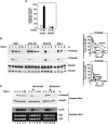
TGF-β induces a prolonged upregulation of snoN expression in AKR-2B and NRK fibroblasts at the level of transcription. (A) TGF-β induces snoN gene expression in multiple cell types. RIE-1, NIH 3T3, NRK, and AKR-2B cells were serum starved for 16 h and stimulated with 100 pM TGF-β1 for various periods of time as indicated. Total RNA was isolated from these cells, and snoN expression was analyzed by Northern blotting. 28S and 18S RNAs are shown as loading controls. Quantification of the Northern blot was carried out using Image J. The density of the snoN signal was normalized to the density of 18S RNA, and the induction of snoN expression by TGF-β in each cell line is shown in the graph. (B) TGF-β upregulates SnoN protein expression in AKR-2B cells. AKR-2B cells were serum starved for 20 h and stimulated with 100 pM TGF-β1 for various amounts of time as indicated. Nuclear extracts were prepared from these cells, and endogenous SnoN was isolated by immunoprecipitation with an anti-SnoN peptide antiserum and analyzed by Western blotting with anti-SnoN (Santa Cruz). As a loading control, nuclear extracts were analyzed by Western blotting with an antibody against nuclear protein HEXIM1. (C) Protein synthesis is not required for TGF-β-induced expression of snoN. AKR-2B cells were treated with or without 100 μg/ml cycloheximide (CHX) in the absence or presence of 100 pM TGF-β1 at the indicated time points. snoN expression was analyzed by Northern blotting. Mouse glyceraldehyde-3-phosphate dehydrogenase (GAPDH) was used as a control for equal loading. (D) TGF-β induces snoN gene expression at the transcriptional level. AKR-2B cells were treated with or without actinomycin D (ActD; 1 μg/ml) in the absence or presence of 100 pM TGF-β1. Fifteen micrograms of total RNA was analyzed by Northern blotting. (E) The prolonged induction of SnoN in AKR-2B cells is not due to reduction of protein degradation. AKR-2B cells were stimulated with TGF-β1 for 2 h followed by treatment with or without 100 μg/ml CHX in the presence of TGF-β1 for another 6 h. Endogenous SnoN was isolated and analyzed as described for panel B. HEXIM1 levels in the nuclear extracts were measured as a loading control. (F) TGF-β induces activation of the snoN promoter. Hep3B cells or AKR-2B cells were transfected with either the sno4.8 reporter plasmid, containing 4.8 kb of the snoN promoter sequence, or the pGL2basic vector control and were stimulated with 50 pM TGF-β1 for 16 h. Luciferase activity was measured 48 h after transfection. (G) Identification of a TGF-β-responsive region in 4.8 kb of the snoN promoter. Hep3B cells were transfected with the luciferase reporter constructs listed in the diagram at the top and were stimulated without (open bars) or with (closed bars) 50 pM TGF-β1 for 16 h.
FIG. 2.
FIG. 2.
Prolonged snoN expression depends on the activation of Smad2. (A) TGF-β-induced snoN expression is Smad dependent. Hep3B cells were transfected with 0.5 μg of sno2.8 alone or together with 2.0 μg of Smad7 and were stimulated without (open bars) or with (closed bars) 50 pM TGF-β for 16 h. (B) Phosphorylation of the R-Smads in response to TGF-β in various cell lines. Cells were treated with 100 pM TGF-β1 for the indicated periods of time. Equal amounts of total-cell lysates prepared from each sample were analyzed by Western blotting using antibodies against phospho-Smad2 (P-Smad2) (top panel), phospho-Smad3 (P-Smad3) (middle panel), or anti-Smad2 and anti-Smad3 (bottom panel). The intensities of phospho-Smad2 and phospho-Smad3 bands in the Western blots were quantified and calculated as percentages of the maximal level of phosphorylation upon TGF-β stimulation (at 2 h) (graphs). Asterisk indicates a nonspecific band recognized by the anti-Smad2 antibody. (C) AKR-2B cells were stimulated with 100 pM TGF-β1 for the indicated times. At 3 h (lanes 8 to 10) or 6 h (lanes 14 and 15) after TGF-β1 addition, dimethyl sulfoxide or 10 μM SB-431542 was added to the culture. Equal amounts of total-cell lysates were analyzed by Western blotting with anti-P-Smad2 or anti-Smad2 (top panels). The levels of snoN mRNA in these cells were analyzed by Northern blotting (bottom panels).
FIG. 3.
FIG. 3.
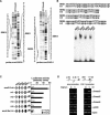
Binding of Smad proteins to the snoN promoter is required for the activation of snoN transcription in response to ΤGF-β1. (A) DNase I footprinting assay. GST-Smad4 was incubated with the indicated 32P-labeled snoN promoter fragments, followed by digestion with DNase I. The regions protected by Smad4 are labeled as SBE1, SBE2, SBE3, and SBE4. The nucleotide sequences of these protected regions were determined by the Maxam-Gilbert G sequencing method. (B) EMSA. (Top) Oligonucleotide probes containing wild-type and mutant Smad binding sites (SBE1 to -4). The WT SBE motifs are capitalized and boldfaced. The mutant sequences are italicized. (Bottom) GST-Smad4 (1 μg) was incubated with 32P-labeled probes in an EMSA. Protein/DNA-binding complexes were visualized by autoradiography. (C) WT or mutant snoN promoter fragments containing mutations in the SBE motifs were cloned into the pGL2 basic vector and transfected into Hep3B cells. Luciferase activity was measured 16 h after TGF-β stimulation. (D) Chromatin immunoprecipitation assay. Soluble chromatin was prepared from Hep3B cells treated with or without TGF-β1 for 1 h. The chromatin immunoprecipitation assay was carried out using various antibodies as indicated. Promoter fragments present in the immunoprecipitates were amplified by PCR using pairs of primers that cover the TGF-β-responsive regions in the snoN and PAI-1 promoters as indicated at the top and detected by agarose gel electrophoresis.
FIG. 4.
FIG. 4.
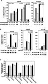
Smad2 and Smad3 play opposite roles in regulation of snoN expression. (A) Hep3B or AKR-2B cells were cotransfected with the indicated Smad complexes and sno2.8 or pLUC800, containing 800 bp of the TGF-β-responsive region from the PAI-1 promoter, in the absence (open bars) or presence (closed bars) of 50 pM TGF-β1 for 16 h. (B) Effects of reduction of Smad2 or Smad3 expression on snoN transcription. Hep3B or AKR-2B cells were transfected with sno2.8 and the pSUPER vector expressing either human Smad2 siRNA, Smad3 siRNAa, a control siRNA (in Hep3B cells), or human Smad3 siRNAb, which cross-reacts with mouse Smad3 (in AKR-2B cells). snoN transcription was examined by the luciferase assay (graphs). The expression levels of Smad2, Smad3, and Smad4 in these luciferase reactions were measured by Western blotting. (C) The DNA binding activity of Smad3 and oligomerization of Smad3 with Smad4 are required for the inhibition of TGF-β-induced snoN transcription. WT Smad4 together with a Smad3 mutant deficient in DNA binding (S3R74A) or defective in hetero-oligomerization (S3V277D), or WT Smad3 together with mutant Smad4 defective in hetero-oligomerization (S4V370D), was cotransfected with sno2.8 into Hep3B cells. As a control, either WT Smad2/Smad4, Smad2(Δexon3)/Smad4, or WT Smad3/Smad4 was cotransfected with sno2.8. Luciferase activity was measured as described for Fig. 1F. Abbreviations: S2/S4, WT full-length Smad2/Smad4; S2(Δexon3)/S4, WT short isoform of Smad2 missing exon3/Smad4; S3/S4, WT Smad3/Smad4 complexes.
FIG. 5.
FIG. 5.
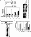
Smad3/Smad4 binds to the Smad-inhibitory element and blocks TGF-β-induced snoN expression. (A) Identification of the SIE. (Top) Internal deletions of the snoN promoter were generated and introduced into Hep3B cells. (Bottom) Luciferase activity was measured in the absence or presence of Smad3/Smad4 and without (open bars) or with (closed bars) 50 pM TGF-β1. (B) Mapping of Smad3 binding site by a DNase I footprinting assay. One microgram of bacterially expressed GST (lane 2), GST-Smad3 (lane 3), or GST-Smad4 (lane 4) was incubated with a 32P-labeled snoN promoter fragment containing the region from kb −2.4 to −2.0, followed by digestion with DNase I. The region protected by Smad3 and Smad4 is shown. (C) EMSA. 32P-labeled oligonucleotide probes containing wild-type or mutant SIE sequences (top) were incubated with 1 μg of GST (lanes 1 and 4), GST-Smad3 (lanes 2 and 5), or GST-Smad4 (lanes 4 and 6) in an EMSA reaction (bottom). (D) SIE mediates inhibition of snoN transcription by Smad3. A luciferase reporter construct containing WT or mutant SIE was transfected into Hep3B cells together with Smad2 and Smad4 (gray bars) or Smad3 and Smad4 (black bars). snoN transcription induced by TGF-β was analyzed by the luciferase assay as described for panel A.
FIG. 6.
FIG. 6.
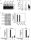
snoN expression is required for TGF-β-induced oncogenic transformation of AKR-2B and NRK cells. (A) Reduction of snoN expression in AKR-2B cells by siRNA. Stable AKR-2B cell lines expressing either the siRNA for murine snoN or an unrelated control siRNA were generated as described in Materials and Methods. The levels of snoN in two representative stable clones (sisnoN#6 and -#13) in the absence or presence of 100 pM TGF-β1 were measured by Northern blotting and compared with levels in parental cells and control siRNA cells. (B) Effects of TGF-β on the growth of parental AKR-2B, control siRNA, and sisnoN cells. A total of 8 × 10⁴ cells were incubated for 4 days in the presence or absence of 100 pM TGF-β1. The growth of cells was quantified by cell counting and normalized to the growth of cells in the absence of TGF-β1. (C) Soft-agar colony assay. A total of 5,000 parental AKR-2B cells or those expressing control siRNA or sisno were subjected to a soft-agar colony assay in the presence of TGF-β1 as described in Materials and Methods. The soft-agar plates were stained with 0.5 mg/ml MTT and scanned. A representative 5-mm2 area of each 6-well plate is shown on the left. The number of soft-agar colonies in each plate was quantified, and the results are summarized in the graph on the right. (D) Prolonged induction of snoN expression by TGF-β is required for the transformation of both AKR-2B and NRK cells. A total of 5,000 AKR-2B cells or 5 × 10⁴ NRK cells were set up in the soft-agar assay as described for panel C. One milliliter of medium with or without 10 μM SB-431542 was added on top of the soft agar at the indicated times post-TGF-β treatment.

References

    1. Attisano, L., and J. L. Wrana. 2002. Signal transduction by the TGF-β superfamily. Science 296:1646-1647. - PubMed
    1. Barnard, J. A., R. M. Lyons, and H. L. Moses. 1990. The cell biology of transforming growth factor β. Biochim. Biophys. Acta 1032:79-87. - PubMed
    1. Bonni, S., H. R. Wang, C. G. Causing, P. Kavsak, S. L. Stroschein, K. Luo, and J. L. Wrana. 2001. TGF-β induces assembly of a Smad2-Smurf2 ubiquitin ligase complex that targets SnoN for degradation. Nat. Cell Biol. 3:587-595. - PubMed
    1. Boyd, K. E., and P. J. Farnham. 1999. Coexamination of site-specific transcription factor binding and promoter activity in living cells. Mol. Cell. Biol. 19:8393-8399. - VSports app下载 - PMC - PubMed
    1. Boyer, P. L., C. Colmenares, E. Stavnezer, and S. H. Hughes. 1993. Sequence and biological activity of chicken snoN cDNA clones. Oncogene 8:457-466. - PubMed

Publication types

MeSH terms

LinkOut - more resources