"VSports在线直播" Skip to main page content
U.S. flag

An official website of the United States government

Dot gov

The . gov means it’s official. Federal government websites often end in . gov or . mil VSports app下载. Before sharing sensitive information, make sure you’re on a federal government site. .

Https

The site is secure. The https:// ensures that you are connecting to the official website and that any information you provide is encrypted and transmitted securely V体育官网. .

. 2005 Nov;37(11):1264-9.
doi: 10.1038/ng1658. Epub 2005 Oct 16.

Identification of a ferrireductase required for efficient transferrin-dependent iron uptake in erythroid cells

Affiliations

"V体育官网" Identification of a ferrireductase required for efficient transferrin-dependent iron uptake in erythroid cells

Robert S Ohgami et al. Nat Genet. 2005 Nov.

Abstract (VSports app下载)

The reduction of iron is an essential step in the transferrin (Tf) cycle, which is the dominant pathway for iron uptake by red blood cell precursors. A deficiency in iron acquisition by red blood cells leads to hypochromic, microcytic anemia. Using a positional cloning strategy, we identified a gene, six-transmembrane epithelial antigen of the prostate 3 (Steap3), responsible for the iron deficiency anemia in the mouse mutant nm1054. Steap3 is expressed highly in hematopoietic tissues, colocalizes with the Tf cycle endosome and facilitates Tf-bound iron uptake. Steap3 shares homology with F(420)H(2):NADP(+) oxidoreductases found in archaea and bacteria, as well as with the yeast FRE family of metalloreductases. Overexpression of Steap3 stimulates the reduction of iron, and mice lacking Steap3 are deficient in erythroid ferrireductase activity VSports手机版. Taken together, these findings indicate that Steap3 is an endosomal ferrireductase required for efficient Tf-dependent iron uptake in erythroid cells. .


Figures
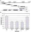
Figure 1
Figure 1
nm1054 physical map and BAC complementation. (a) Physical map of the nm1054 region demonstrating the extent of the deletion interval (shaded region) based on sequences in the Ensembl database (www.ensembl.org; April 2005 release). Microsatellite markers (top), BAC transgenes (middle), and genes (bottom) are shown. CITB and RPCI-22 BAC transgenes are indicated by their clone ID numbers. Partial insertions of RPCI-22 11D19 are indicated by an asterix (*). Ensembl transcript ENSMUST00000037840; Sctr, secretin receptor; Pr1, Neuronal voltage-gated calcium channel γ-like subunit; Dbi, diazepam binding inhibitor; RefSeq_dna ID NM_028439; Steap3, six-transmembrane epithelial antigen of the prostate 3; C1ql2, complement component 1, q subcomponent 2-like (b) Comparison of hemoglobin (HGB [g/l]) levels in 4 week old BAC transgenic [C57BL/6 x 129S6/SvEvTac] F1 animals. ?/+ includes all nm/+ and +/+ animals with or without a BAC transgene (n=12). Other groups are nm/nm animals without a BAC transgene (Tg; n=11), a transgene other than RPCI-22 11D19 (Tg+; n=15), a partial insertion of RPCI-22 11D19 (Tg11D19*+; n=6), or a complete insertion of RPCI-22 11D19 (Tg11D19+). Error bars ± 1 standard deviation (S.D.).
Figure 2
Figure 2
Steap3 mRNA expression (a) In situ hybridization of E15.5 mouse embryo demonstrating high-level fetal liver (open arrow) and labyrinthine placental expression (closed arrow). (b) Quantitative real time PCR of STEAP3 in human tissues. Relative RNA abundance is normalized to spleen, which was defined as a ratio of 1.0. Error bars ± 1 S.D.
Figure 3
Figure 3
Steap3 sub-cellular localization. (a–i) Co-localization of epitope-tagged Steap3 with endogenous Tf and Tfr1, and epitope-tagged DMT1. (a) Steap3, (b) Tf, (c) Tf-Steap3 merged; (d) Steap3, (e) Tfr1, (f) Tfr1-Steap3 merged, (g) Steap3, (h) Dmt1, (i) Dmt1-Steap3 merged.
Figure 4
Figure 4
Somatic complementation of the nm1054 anemia in bone marrow chimeras. (a,b) Comparison of peripheral blood smears of lethally irradiated wild type recipient mice transplanted with homozygous mutant fetal liver hematopoietic cells transduced with (a) the retroviral vector alone, or (b) a construct expressing Steap3. (c) Hematological data and RBC chimerism in transplant chimeras at 4 weeks post-transplantation (n=4 in each group). HGB=hemoglobin, MCV=mean RBC volume, ZPP/Heme=RBC zinc protoporphyrin IX: iron protoporphyrin IX (heme) ratio. Error bars ± 1 S.D. Student’s T-test P= 0.009 (HGB), 0.017 (MCV), <0.001 (ZPP/Heme). Chimerism was not significantly different between the two groups (P=0.837). Error bars ± 1 S.D.
Figure 5
Figure 5
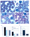
Hematologic data from nm1054 and Steap3 null mice. (a–d) Peripheral blood smears of (a) Steap3+/+, (b) Steap3nm1054/nm1054, (c) Steap3nm1054/, and (d) Steap3−/− animals at 8 weeks of age. (e) Hematological data in 8 week old Steap3+/+ (n=5), Steap3nm1054/nm1054 (n=6), Steap3nm/1054/ (n=6), and Steap3−/− (n=6) mice. Error bars ± 1 S.D. See also Supplementary Table 1 online. (f) Iron uptake activity in Steap3+/+ (n=5) and Steap3−/− (n=6) reticulocyte-rich RBCs. The fraction of the total iron, present as heme, is indicated in the filled portion of the bar.
Figure 6
Figure 6
Structure of the Steap family and function of Steap3 (a) Schematic diagram of Steap1-4 and yeast FRE1. Blue ovals in tandem represent the unique flavin- NAD(P)H binding domain. Heme groups are indicated in red. The green in FRE1 represents the FAD:NAD(P)H domain. (b) (top) Multiple sequence alignment of the flavin F420H2: NADP+ Oxidoreductase (FNO) from Archaeoglobus fulgidus and the Mus musculus Steap family members. Steap lacks the FNO-like domain. Conserved residues in the Rossman fold motif (GXGXXA/G) are in bold italics. Serine (S) and arginine (R) residues critical for binding to the phosphate moiety of NADP+ are in bold. (bottom) Predicted transmembrane domains 3 (TM3) and 5 (TM5) of the Steap proteins are aligned with Saccharomyces cerevisiae FRE1. Conserved histidines (H) residues are in bold. The second, non-conserved histidine in each FRE1 TM is in bold italic. Multiple sequence alignment was performed with MultAlign (http://cbrg.inf.ethz.ch/Server/MultAlign.html). (c) Ferrireductase activity in Steap3+/+ (n=10) and Steap3nm1054/nm1054 (n=5) reticulocyte-rich RBCs (d) Ferrireductase activity in Steap3+/+ (n=6) and Steap3−/− (n=5) reticulocyte-rich RBCs. (e) Ferrireductase activity in HEK 293T cells transfected with vector alone (n=12), or wild type (n=12), or mutant Steap3 (n=6 each) expression constructs. Error bars ± 1 S.D. (f) Schematic model of Steap3 mediated reduction of iron. Putative Steap3 membrane topology, FNO motif, and heme coordination sites located in TM3 and TM5.

Comment in

References

    1. Ponka P. Tissue-specific regulation of iron metabolism and heme synthesis: distinct control mechanisms in erythroid cells. Blood. 1997;89:1–25. - PubMed
    1. Fleming MD, et al. Microcytic anemia mice have a mutation in Nramp2, a candidate iron transporter gene. Nat Genet. 1997;16:383–386. - PubMed
    1. Fleming MD, et al. Nramp2 is mutated in the anemic Belgrade (b) rat: evidence of a role for Nramp2 in endosomal iron transport. Proc Natl Acad Sci U S A. 1998;95:1148–53. - PMC - PubMed
    1. Gunshin H, et al. Cloning and characterization of a mammalian proton-coupled metal-ion transporter. Nature. 1997;388:482–488. - PubMed
    1. McKie AT, et al. An iron-regulated ferric reductase associated with the absorption of dietary iron. Science. 2001;291:1755–1759. - PubMed

Publication types

MeSH terms