"V体育官网入口" Skip to main page content
U.S. flag

An official website of the United States government

Dot gov

The . gov means it’s official. Federal government websites often end in VSports app下载. gov or . mil. Before sharing sensitive information, make sure you’re on a federal government site. .

Https

The site is secure. The https:// ensures that you are connecting to the official website and that any information you provide is encrypted and transmitted securely V体育官网. .

Comparative Study
. 2005 Oct 17;202(8):1063-73.
doi: 10.1084/jem.20051100. Epub 2005 Oct 10.

Functional specialization of gut CD103+ dendritic cells in the regulation of tissue-selective T cell homing

Affiliations
Comparative Study

VSports最新版本 - Functional specialization of gut CD103+ dendritic cells in the regulation of tissue-selective T cell homing

Bengt Johansson-Lindbom et al. J Exp Med. .

Abstract

Gut-associated lymphoid tissue (GALT) dendritic cells (DCs) display a unique ability to generate CCR9+alpha4beta7+ gut-tropic CD8+ effector T cells. We demonstrate efficient induction of CCR9 and alpha4beta7 on CD8+ T cells in mesenteric lymph nodes (MLNs) after oral but not intraperitoneal (i VSports手机版. p. ) antigen administration indicating differential targeting of DCs via the oral route. In vitro, lamina propria (LP)-derived DCs were more potent than MLN or Peyer's patch DCs in their ability to generate CCR9+alpha4beta7+ CD8+ T cells. The integrin alpha chain CD103 (alphaE) was expressed on almost all LP DCs, a subset of MLN DCs, but on few splenic DCs. CD103+ MLN DCs were reduced in number in CCR7-/- mice and, although CD8+ T cells proliferated in the MLNs of CCR7-/- mice after i. p. but not oral antigen administration, they failed to express CCR9 and had reduced levels of alpha4beta7. Strikingly, although CD103+ and CD103- MLN DCs were equally potent at inducing CD8+ T cell proliferation and IFN-gamma production, only CD103+ DCs were capable of generating gut-tropic CD8+ effector T cells in vitro. Collectively, these results demonstrate a unique function for LP-derived CD103+ MLN DCs in the generation of gut-tropic effector T cells. .


VSports手机版 - Figures

Figure 1.
Figure 1.
Efficient and adjuvant-independent generation of CCR9+ α4β7+ gut-homing T cells in the MLNs after oral administration of antigen. After adoptive transfer of OT-I cells (A–C) or an equal number of CCR9−/− and WT OT-I cells (D), recipient mice were injected i.p. with 5 mg OVA ± 50 μg LPS or given 50 mg OVA ± 20 μg CT orally. 3 d later, donor cells in MLNs (A–C) or MLNs and the small intestinal epithelium (D) were analyzed by flow cytometry. (A and B) Expression of CCR9 and α4β7 by OT-I cells in MLNs. Numbers indicate the percentages of positive cells. One representative experiment of between five and seven performed. (C) Pooled results from mice receiving OVA alone orally (shaded bars) or i.p. (open bars). Mean ± SD of between five and seven experiments with three to four mice in each experiment. *, P < 0.05; **, P < 0.005. (D) Ratio of CCR9−/− to WT OT-I cells in the MLNs and IEL compartment. Mean ± SD of three separate experiments with three mice per group in each experiment.
Figure 2.
Figure 2.
LP dendritic cells are potent in generating CCR9+ α4β7+ CD8+ T cells. OT-I cells were activated in vitro with SIINFEKL peptide–pulsed DCs purified from LP, MLNs, PP, or spleen. (A) OT-I cell proliferation in response to a graded number of indicated DCs as assessed by quantification of methyl-[3H]thymidine incorporation. Values represent mean ± SD. (B) CCR9 and (C) α4β7 expression by CFSE-labeled OT-I cells was determined by flow cytometry after 4–5 d of co-culture with DCs. The percentage of positive cells among dividing OT-I cells is presented. DCs from Flt3L-treated mice were used in all experiments except experiment (expt) 4 in (B) and (C), where DCs were purified from pooled tissues of 10 untreated mice. *, P < 0.05. SPL, spleen.
Figure 3.
Figure 3.
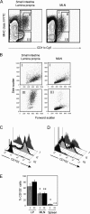
CD103 is expressed by the majority of LP DCs and a subset of MLN DCs. Leukocytes were isolated from the small intestinal LP, MLNs, and spleen and analyzed by flow cytometry using 7-AAD, anti–MHC class II–FITC, anti-CD11c–APC, and anti-CD103–PE mAbs. (A) Identification of CD11c+MHC class II+ (region I), CD11chighMHC class II+ (region II), and CD11clowMHC class II (region III) cells after gating on 7-AAD (live) cells. (B) Light scatter properties of the indicated populations of cells. (C–D) Representative histograms showing CD103 expression by LP cells (C) and MLN cells (D) using the region definitions depicted in (A). (E) Statistical analysis of CD103 expression by CD11c+MHC class II+ (region I) and CD11chighMHC class II+ (region II) DCs from LP, MLNs, and spleen (mean ± SD; n = 9 for LP and MLNs, n = 6 for spleen). *, P < 0.0005; **, P < 0.0001 compared with LP equivalent. n.d., not done.
Figure 4.
Figure 4.
In situ expression of CD103 by LP DCs. Cryostat sections of the small intestinal jejunum of 8-wk-old C57BL/6 mice were analyzed for expression of CD11c, MHC class II, and CD103 by four-color immunofluorescence using DAPI for visualization of nuclei. Images show overlays of CD11c (green) and DAPI (blue) either alone (A and D) or in combination with CD103 (red; B and E) or MHC class II (red; C and F). CD11c+MHC class II+ DCs coexpressing CD103 (arrows) were found in the LP of the villi either as scattered cells (A–C) or in clusters (D–F). The contrast and γ parameters have been modified for CD11c- and MHC class II–derived fluorescence in order to balance the high intensity of the CD103 staining.
Figure 5.
Figure 5.
Phenotype of CD103+ and CD103 DCs in the small intestinal LP and MLNs. Leukocytes were isolated from the small intestinal LP and MLNs, incubated with Cy5-conjugated anti-CD11c mAb, 7-AAD, and mAbs against the indicated proteins, and analyzed by flow cytometry. Expression levels of these proteins are shown after gating on 7-AAD (live) and CD11c+ (MLN) or CD11chigh (LP) cells. Because the flow cytometer was equipped with four photomultiplier tubes for fluorescence only, we could not include an MHC class II staining in these analyses. CD11clow LP cells were therefore not considered because these include the MHC class II and granular cells contained within region III that is shown in Fig. 3 A. The numbers indicate mean fluorescence intensity ± SD, except for the CD62L analysis, which indicates the percentage of cells in each quadrant.
Figure 6.
Figure 6.
CD8+ T cells primed in the MLNs of CCR7/ mice fail to adopt a gut-tropic phenotype. CFSE-labeled OT-I cells were adoptively transferred into WT or CCR7−/− recipient mice. 3 d after i.p. immunization with OVA and LPS, MLNs were collected, and the phenotype of OT-I cells was determined by flow cytometry. (A) Representative data of CCR9, α4β7, and CD62L expression by divided OT-I cells in the MLNs of WT versus CCR7−/− recipient mice. The CFSE gate is set to distinguish dividing from nondividing cells and is based on the CFSE intensity of OT-I cells in the PLNs of recipient mice (that do not divide) 3 d after oral OVA administration. The percentages indicate divided cells that express the markers shown. (B) Mean values ± SD obtained with three mice in each group.
Figure 7.
Figure 7.
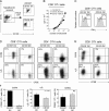
CD103+ but not CD103 MLN DCs can generate gut-tropic CCR9+ α4β7high T cells. (A) CD11c+ DCs were enriched from MLNs using anti-CD11c magnetic bead cell sorting, incubated with fluorescently labeled mAbs against CD11c and CD103, and sorted into CD103+ and CD103 DCs by FACS. (B–F) Indicated DC subsets were loaded with OVA peptide and co-cultured with CD8+ OT-I or CD4+ OT-II cells. (B) Proliferation of OT-I cells responding to a graded number of DCs was determined by methyl-[3H]thymidine incorporation. One representative experiment of three performed is shown. (C) IFN-γ production by OT-I cells was determined by flow cytometry after expansion with IL-7 and IL-15. Percentages shown indicate OT-I cells expressing IFN-γ. (D–F) CFSE-labeled OT-I or OT-II cells were cultured with CD103+ or CD103 DCs for 4–5 d (D) and further expanded in IL-7 and IL-15 for 3 d (E). Expression of CCR9, α4β7, and CD62L by responding T cells was then analyzed by flow cytometry. (D and E) Results are from one representative experiment of four (OT-I) and two (OT-II) performed. The percentages indicate divided cells expressing the indicated markers. (F) Mean ± SD from four separate experiments in which expression of CCR9 and CD62L was analyzed after the primary DC culture, and α4β7 was analyzed after further expansion in IL-7 and IL-15.

Comment in

References

    1. Kunkel, E.J., and E.C. Butcher. 2002. Chemokines and the tissue-specific migration of lymphocytes. Immunity. 16:1–4. - PubMed
    1. Campbell, D.J., and E.C. Butcher. 2002. Rapid acquisition of tissue-specific homing phenotypes by CD4+ T cells activated in cutaneous or mucosal lymphoid tissues. J. Exp. Med. 195:135–141. - PMC - PubMed
    1. Svensson, M., J. Marsal, A. Ericsson, L. Carramolino, T. Broden, G. Marquez, and W.W. Agace. 2002. CCL25 mediates the localization of recently activated CD8alphabeta(+) lymphocytes to the small-intestinal mucosa. J. Clin. Invest. 110:1113–1121. - V体育安卓版 - PMC - PubMed
    1. Calzascia, T., F. Masson, W. Di Berardino-Besson, E. Contassot, R. Wilmotte, M. Aurrand-Lions, C. Ruegg, P.Y. Dietrich, and P.R. Walker. 2005. Homing phenotypes of tumor-specific CD8 T cells are predetermined at the tumor site by crosspresenting APCs. Immunity. 22:175–184. - PubMed
    1. Johansson-Lindbom, B., M. Svensson, M.-A. Wurbel, B. Malissen, G. Márquez, and W. Agace. 2003. Selective generation of gut-tropic T cells in gut-associated lymphoid tissue (GALT); requirement for GALT dendritic cells and adjuvant. J. Exp. Med. 198:963–969. - PMC - PubMed

Publication types

MeSH terms