VSports - Skip to main page content
U.S. flag

An official website of the United States government

Dot gov

The . gov means it’s official. Federal government websites often end in . gov or . mil. Before sharing sensitive information, make sure you’re on a federal government site. VSports app下载.

Https

The site is secure. The https:// ensures that you are connecting to the official website and that any information you provide is encrypted and transmitted securely. V体育官网.

Comparative Study
. 2005 Sep 23;122(6):875-86.
doi: 10.1016/j.cell.2005.07.012.

General translational repression by activators of mRNA decapping

Affiliations
Comparative Study

General translational repression by activators of mRNA decapping

Jeff Coller et al. Cell. .

Abstract

Translation and mRNA degradation are affected by a key transition where eukaryotic mRNAs exit translation and assemble an mRNP state that accumulates into processing bodies (P bodies), cytoplasmic sites of mRNA degradation containing non-translating mRNAs, and mRNA degradation machinery. We identify the decapping activators Dhh1p and Pat1p as functioning as translational repressors and facilitators of P body formation. Strains lacking both Dhh1p and Pat1p show strong defects in mRNA decapping and P body formation and are blocked in translational repression. Contrastingly, overexpression of Dhh1p or Pat1p causes translational repression, P body formation, and arrests cell growth. Dhh1p, and its human homolog, RCK/p54, repress translation in vitro, and Dhh1p function is bypassed in vivo by inhibition of translational initiation. These results identify a broadly acting mechanism of translational repression that targets mRNAs for decapping and functions in translational control VSports手机版. We propose this mechanism is competitively balanced with translation, and shifting this balance is an important basis of translational control. .


Figures

Figure 1
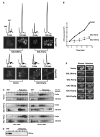
Figure 1. Dhh1p and Pat1p Have Additive Effects on P Body Formation and Are Required for Translational Repression
(A) Using Dcp2p-GFP as a marker, P bodies were observed in wild-type (WT), dhh1Δ, pat1Δ, dhh1Δ/pat1Δ, or dcp1Δ strains. (B) Depicted are polysome profiles (OD260nm traces) of WT, dhh1Δ, pat1Δ, dhh1Δ/pat1Δ, or dcp1Δ before and after glucose deprivation. The nontranslating region (RNP), 80S monosome (80S), and polysomes (P) are indicated above. (C) Northern of RPL41a mRNA following polysome analysis in WT or dhh1Δ/pat1Δ before and after glucose deprivation. (D) Incorporation of [35S]methionine in WT and dhh1Δ/pat1Δ cells before and after glucose deprivation. Values are reported as cpm incorporated. Time after label addition indicated. (E) Polysome profiles (OD260nm traces) of WT and dhh1Δ/pat1Δ strains before and after amino acid deprivation.
Figure 2
Figure 2. Overexpression of Dhh1p or Pat1p Causes General Translational Repression and Increases P Bodies
(A) Polysome profiles and P body accumulation in control cells (vector) or cells overexpressing Dhh1p, Pat1p, or Dcp1p. Times after induction by the addition of galactose are indicated. P bodies were visualized using Dcp2p-GFP. (B) Incorporation of [35S]methionine in cells overexpressing Dhh1p, Pat1p, or a vector control. Values are reported as cpm incorporated. Time after addition of label indicated. (C) Northerns of the PGK1 and RPL41a mRNA following polysome analysis, in control cells, or cells overexpressing Dhh1p or Pat1p. Minutes after induction are indicated. Nontranslating (RNP) and translating areas (polysomes) are indicated above. (D) Western analysis indicating location of Dhh1p on a sucrose gradient. (E) Growth of yeast cells when Dhh1p, Pat1p, Dcp1p, Dcp2p, or Edc3p are overexpressed. Cells were plated on either glucose media, or galactose.
Figure 3
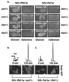
Figure 3. Dhh1p and Pat1p Stimulate Translational Repression via Parallel Pathways
(A) Shown is growth of yeast cells when Dhh1p or Pat1p are overexpressed in strains deleted for components of P bodies. Cells were plated on glucose media or galactose media. (B) Polysome and P body analysis of cells overexpressing Dhh1p in a pat1Δ strain or (C) Pat1p in a dhh1Δ strain. Nontranslating region (RNP), 80S monosome (80S), and polysomes (P) are indicated above first trace. P bodies were visualized using Dcp2p-GFP. Time after galactose induction is indicated.
Figure 4
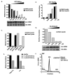
Figure 4. Dhh1p and Its Human Homolog RCK/p54 Stimulate Translational Repression In Vitro
(A) GST-Dhh1p, (B) GST-Ded1p, (C) GST-Dhh1-8p, or (D) His-RCK/p54 was added to either yeast or reticulocyte extracts. LUC activity normalized to translation observed in absence of test protein. Protein concentration is indicated below graph. For (C), 7.3 μM of protein was added. Northern of LUC reporter from extracts containing Dhh1p (A) or RCK/p54 (D). (E) Graph indicates relative LUC activity of cap plus (Cap+Luc) and cap minus (Luc) LUC reporter in the presence of buffer, 1.0 μM GST-Dhh1p, or 2.0 μM GST-Dhh1p. (F) Dhh1p inhibited 48S preinitiation complex formation in vitro. Extracts were programmed with radiolabeled reporter and GMPPNP in the presence or absence of 7.3 μM GST-Dhh1p. Graph indicates percent of radiolabeled reporter in each fraction. Position of the RNP, 48S, and 80S complexes are indicated.
Figure 5
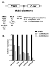
Figure 5. General Translational Inhibition by RCK/p54
LUC activity of various IRES-containing mRNAs in rabbit reticulocyte lysate with 1 μM, 2 μM, or no recombinant RCK/p54. Activity is normalized to translation in absence of RCK/p54. Identity of each construct is indicated below graph, and corresponds to (A).
Figure 6
Figure 6. Dhh1p Function Requires Translational Initiation In Vivo
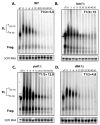
Decay of SL-PGK1 reporter in (A) WT, (B) lsm1Δ, (C) pat1Δ, and (D) dhh1Δ strains. Time points are indicated above each lane. Half-lives are indicated in minutes. Diagram indicates position of the full-length mRNA and polyguanosine decay intermediate. Poly(A) tail lengths are determined by comparing mRNA size to a oligo dT/RNaseH treated control (dT).
Figure 7
Figure 7. A General Active Repression Machinery Exists that Is in Competition with Translation
This competition creates a finely balanced system setting the relative translation rate for an mRNA. mRNAs can be driven into either translation or repression by tipping the balance of this competition via any number of events.

References

    1. Akao Y, Mizoguchi H, Ohishi N, Yagi K. Growth inhibition by overexpression of human DEAD box protein rck/p54 in cells of a guinea pig cell line. FEBS Lett. 1998;429:279–283. - VSports最新版本 - PubMed
    1. Andrei M, Ingelfinger D, Heintzmann R, Achsel T, Rivera-Pomar R, Luhrmann R. A role for eIF4E and eIF4E-transporter in targeting mRNPs to mammalian processing bodies. RNA. 2005;11:717–727. - "VSports注册入口" PMC - PubMed
    1. Ashe M, De Long S, Sachs A. Glucose depletion rapidly inhibits translation initiation in yeast. Mol Biol Cell. 2000;11:833–848. - PMC - PubMed
    1. Bashkirov V, Scherthan H, Solinger J, Buerstedde J, Heyer W. A mouse cytoplasmic exoribonuclease (mXRN1p) with preference for G4 tetraplex substrates. J Cell Biol. 1997;136:761–773. - PMC - PubMed
    1. Bonnerot C, Boeck R, Lapeyre B. The two proteins Pat1p (Mrt1p) and Spb8p interact in vivo, are required for mRNA decay, and are functionally linked to Pab1p. Mol Cell Biol. 2000;16:5939–5946. - PMC - PubMed

VSports - Publication types

MeSH terms

Substances