VSports在线直播 - Skip to main page content
U.S. flag

An official website of the United States government

Dot gov

The . gov means it’s official. Federal government websites often end in . gov or . mil. Before sharing sensitive information, make sure you’re on a federal government site VSports app下载. .

Https

The site is secure V体育官网. The https:// ensures that you are connecting to the official website and that any information you provide is encrypted and transmitted securely. .

. 2005 Sep;116(1):21-9.
doi: 10.1111/j.1365-2567.2005.02192.x.

Detection, epitope-mapping and function of anti-Fas autoantibody in patients with silicosis

Affiliations

VSports最新版本 - Detection, epitope-mapping and function of anti-Fas autoantibody in patients with silicosis

"VSports" Akiko Takata-Tomokuni et al. Immunology. 2005 Sep.

Abstract

Dysregulation of apoptosis through the Fas-Fas ligand pathway is associated with the onset of autoimmune disease. Since autoantibodies directed against unknown antigens are present in the sera of these patients, sera samples were examined for the presence of autoantibodies directed against the Fas molecule. Using Western blotting and a ProteinChip analysis, autoantibodies against Fas were detected in patients with silicosis, systemic lupus erythematosus (SLE) and systemic sclerosis (SSc), and weakly detected in healthy individuals. Using epitope mapping employing 12-amino-acid polypeptides with the SPOTs system, a minimum of four epitopes and a maximum of 10 epitopes were found. Several amino acid residues involved in binding FasL, such as C66, R87, L90, E93 and H126, were presented within the epitopes. Serum containing a large amount of anti-Fas autoantibody from silicosis patients inhibited the growth of a Fas-expressing human cell line, but did not inhibit the growth of a low Fas-expresser nor a Fas-expresser in which the Fas gene had been silenced by small interference RNA. All epitopes in the intracellular region of Fas were located in the death domain. The possible roles of anti-Fas autoantibody detected in healthy volunteers and patients with silicosis or autoimmune diseases are discussed here. VSports手机版.


Figures
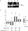
Figure 1
Figure 1
Detection of anti-Fas autoantibodies by Western blot analysis. (a) Human Fas linked to glutathione-s-transferase was separated by 10% sodium dodecyl sulphate–polyacrylamide gel electrophoresis and electrotransferred to a polyvinylidene difluoride membrane. The blots were incubated with sera (1/100 dilution) from either a healthy volunteer (HV) or patients with silicosis (SIL), systemic lupus erythematosus (SLE) or systemic sclerosis (SSc). The blot was then incubated with horseradish peroxidase-conjugated sheep anti-human immunoglobulin (1/15 000 dilution). The bound antibodies were detected using the enhanced chemiluminescence method. (b) The ratio of the intensity of the detected band in the patient samples was calculated relative to that of a healthy volunteer (HV) analysed at the same time. The cut-off point was determined to be 1·2 by the receiver operating characteristic curve (indicated by the thick dotted line). The percentage of positive sera for each patient is also shown. Open circles represent outlier values.
Figure 2
Figure 2
Surface enhanced laser desorption/ionization (SELDI) ProteinChip analysis of anti-Fas autoantibodies. (a) Human Fas antigen (450 ng/2 μl/spot) was loaded on the spots of a preactivated SELDI ProteinChip and incubated at room temperature for 1 hr. The chip was blocked with 1 m ethanolamine and incubated with rabbit anti-human Fas polyclonal antibody as a positive control (top panel) overnight at 4°. The whole chip array was treated with saturated sinapinic acid (an energy absorbing molecule), and analysed using the SELDI ProteinChip System. To clarify whether non-specific binding is found under the same conditions, another chip was treated with PBS (without Fas antigen) and serum of a healthy volunteer (HV-1) (middle panel), or with Fas antigen and PBS (bottom panel). (b) ProteinChip arrays were treated in the same way as in (a), except that the arrays were incubated with human sera instead of rabbit antibody. The x-axes in each figure indicate a molecular weight of 130 000–170 000, and the y-axes show the relative density of the arrays. The molecular weight of the antibody in serum was determined from the peak of the reacted density. Antibodies of approximately 148 000 MW in eight serum samples (SIL-1, SIL-2, SLE-1, SLE-2, SSc-1 and HV-1, HV-2, HV-3), 152 000 in three serum samples (SIL-3, SIL-4 and SSc-2), and two molecular weight antibodies, 148 000 and 152 000, in two serum samples (SLE-3 and SSc-3) were found. (c) The integrated value of the 130 000–170 000 peak area for each sample was calculated from the graph obtained in (b) and was normalized against that of the positive control.
Figure 3
Figure 3
Epitope mapping of autoantibodies directed against human Fas. Oligopeptides were synthesized on activated membranes using the SPOTs system. The peptides were 12 amino acids in length and had a sequential overlap of nine amino acids. The membranes were blocked, incubated with 1/50 diluted sample sera, then detected using β-galactosidase-conjugated goat anti-human immunoglobulin antibody. Hatched sequences indicate each epitope recognized by the autoantibodies.
Figure 4
Figure 4
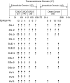
The location of each epitope on the Fas molecule recognized by the anti-Fas autoantibodies in sera from patients with silicosis (SIL), systemic lupus erythematosus (SLE), systemic sclerosis (SSc) and healthy volunteers (HV). The numbers with and without parentheses on the Fas molecule indicate the number of amino acids within the specified domains and the amino acid positioning of the domains within the Fas molecule, respectively. CRD, cysteine-rich domain.
Figure 5
Figure 5
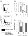
(a) Surface Fas expression levels of KMS-12PE and KMS-12BM human myeloma sister cell lines. The filled histogram indicates the cell only control and the open histogram indicates cells stained with fluorescein isothiocyanate-labelled anti-Fas monocloal antibody. (b) The relative growth of KMS-12PE and KMS-12BM cells cultured with RPMI-1640 medium plus 5% FBS with or without (control) the Fas-mediated apoptosis inducing CH11 antibody (50 and 100 ng/ml) as analysed by the WST-1 assay with the control value being set at 1·0. Although growth of the Fas-expressing KMS-12PE cell line was inhibited by CH11, growth of KMS-12BM cells showed no change. (c) KMS-12PE and KMS-12BM cell lines were cultured with serum from HV-6 or SIL-1 (the SIL-1 serum contained a large amount of anti-Fas autoantibodies as analysed by Western blotting and ProteinChip System). The serum from SIL-1 inhibited growth of KMS-12PE, a Fas-expresser, but not KMS-12BM, a low Fas-expresser.
Figure 6
Figure 6
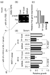
(a) Time schedule of siRNA experiments. Details are given in the Materials and methods. (b) The gel image of MP-RT-PCR for the Fas gene in KMS-12PE cells cultured for −24–0 hr with RPMI-1640 medium supplemented with 5% fetal bovine serum with (TF Cont) or without (Cont) transfection control medium or siRNA medium. (c) The relative Fas gene expression level was analysed from the gel-image shown in (b). Although the TF Cont showed a slight reduction, the siRNA sample demonstrated a marked decrease in Fas gene expression. (d) After harvesting RNA samples, each group of cells was resuspended in the appropriate medium containing 5% serum from either HV-6 or SIL-1. After 24 and 48 hr, the WST-1 assay was performed. At both time points, silencing the Fas gene led to a statistically significant recovery of the SIL-1 serum-induced growth inhibition.

References

    1. Tomokuni A, Aikoh T, Matsuki T, et al. Elevated soluble Fas/APO-1 (CD95) levels in silicosis patients without clinical symptoms of autoimmune diseases or malignant tumours. Clin Exp Immunol. 1997;110:303. - PMC - PubMed
    1. Otsuki T, Sakaguchi H, Tomokuni A, et al. Soluble Fas mRNA is dominantly expressed in cases with silicosis. Immunology. 1998;94:258. 10.1046/j.1365-2567.1998.00509.x. - DOI - PMC - PubMed
    1. Tomokuni A, Otsuki T, Sakaguchi H, Isozaki Y, Hyodoh F, Kusaka M, Ueki A. Detection of anti-topoisomerase I autoantibody in patients with silicosis. Environ Health Prev Med. 2002;7:7. V体育2025版 - 10.1265/ehpm.2002.7. - DOI - PMC - PubMed
    1. Ueki H, Kohda M, Nobutoh T, et al. Antidesmoglein autoantibodies in silicosis patients with no bullous diseases. Dermatology. 2001;202:16. 10.1159/000051578. - DOI - PubMed
    1. Ueki A, Isozaki Y, Tomokuni A, et al. Intramolecular epitope spreading among anti-caspase-8 autoantibodies in patients with silicosis, systemic sclerosis and systemic lupus erythematosus, as well as in healthy individuals. Clin Exp Immunol. 2002;129:556. VSports注册入口 - 10.1046/j.1365-2249.2002.01939.x. - DOI - PMC - PubMed

MeSH terms