Skip to main page content
U.S. flag

An official website of the United States government

Dot gov

The . gov means it’s official. Federal government websites often end in . gov or . mil. Before sharing sensitive information, make sure you’re on a federal government site. VSports app下载.

Https

The site is secure V体育官网. The https:// ensures that you are connecting to the official website and that any information you provide is encrypted and transmitted securely. .

. 2005 Aug 15;202(4):485-92.
doi: 10.1084/jem.20050456. Epub 2005 Aug 8.

Characterization of the early stages of thymic NKT cell development

Affiliations

VSports在线直播 - Characterization of the early stages of thymic NKT cell development

Kamel Benlagha et al. J Exp Med. .

"V体育平台登录" Abstract

Upon reaching the mature heat stable antigen (HSA)low thymic developmental stage, CD1d-restricted Valpha14-Jalpha18 thymocytes undergo a well-characterized sequence of expansion and differentiation steps that lead to the peripheral interleukin-4/interferon-gamma-producing NKT phenotype. However, their more immature HSAhigh precursors have remained elusive, and it has been difficult to determine unambiguously whether NKT cells originate from a CD4+ CD8+ double-positive (DP) stage, and when the CD4+ and CD4- CD8- double-negative (DN) NKT subsets are formed. Here, we have used a CD1d tetramer-based enrichment strategy to physically identify HSAhigh precursors in thymuses of newborn mice, including an elusive DPlow stage and a CD4+ stage, which were present at a frequency of approximately 10(-6) VSports手机版. These HSAhigh DP and CD4+ stages appeared to be nondividing, and already exhibited the same Vbeta8 bias that characterizes mature NKT cells. This implied that the massive expansion of NKT cells is separated temporally from positive selection, but faithfully amplifies the selected TCR repertoire. Furthermore, we found that, unlike the DN gammadelta T cells, the DN NKT cells did not originate from a pTalpha-independent pathway bypassing the DP stage, but instead were produced during a short window of time from the conversion of a fraction of HSAlow NK1. 1neg CD4 cells. These findings identify the HSAhigh CD4+ stage as a potential branchpoint between NKT and conventional T lineages and between the CD4 and DN NKT sublineages. .


Figures

Figure 1.
Figure 1.
Detection of HSAhigh NKT lineage cells in total thymocyte preparations. Total thymocytes from 2-wk-old WT, Jα18−/−, and CD1d−/− mice were stained with HSA and CD1d-tetramers. Gates identifying the tet+HSAlow and tet+HSAhigh cells are shown with corresponding frequencies. Note that the level of background staining in the HSAhigh gates (as measured in Jα18−/− and CD1d−/− mice) precludes the identification of HSAhigh NKT lineage cells in wild type mice. Data representative of >10 individual WT or mutant mice.
Figure 2.
Figure 2.
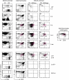
Improved detection of HSAhigh NKT lineage cells in MACS-enriched thymocyte preparations. Thymocytes from 3-d-old WT, Jα18−/−, and CD1d−/− mice were MACS-enriched for tet+ cells and stained for HSA, CD4, and CD8. The left column shows CD1d-tetramer/HSA staining of MACS-enriched preparations, the other columns show CD4/CD8 staining of whole thymocytes, or MACS-enriched thymocytes in the R1 (HSAlow) and R2 (HSAhigh) gates, as indicated. The numbers on the HSA/CD1d-tetramers dot plots represent the percentages of HSAlow and HSAhigh cells among the tet+ population. The numbers on the R1 and R2 gated CD4/CD8 FACS dot plots represent the percentage of CD4+ and DN populations among HSAlow and HSAhigh NKT lineage cells. Note that in the tet+ MACS-enriched panels, each dot represents a cell: there were 3 and 5 tet+ dots in the MACS-enriched preparations of Jα18−/− and CD1d−/− thymuses, respectively, and 224 tet+ dots in the WT. Data are representative of at least three individual experiments with pooled thymuses from 40 to 60 3-d-old newborns.
Figure 3.
Figure 3.
Ontogeny of HSAhigh and HSAlow NKT lineage thymocytes. Thymocytes of WT mice at different ages were enriched and analyzed as in Fig. 2. The total numbers of MACS-enriched tet+ cells at 4 and 5 d was 208 and 265, respectively (note that all dots are displayed and each dot represents a cell). Tet+ DPlow cells are in the pink elliptic gate and have the same DPlow phenotype as the CD69+ DP cells in the total thymocyte population shown in the rightmost panel of fourth row. Note that for consistency of CD4/CD8 staining, thymuses at 8 d, 14 d, and 5 weeks were analyzed side by side in the same experiment (rows 3–5), with doublet and propidium iodide uptake exclusion. The 2-wk-old thymocytes also were analyzed side by side with Jα18−/− and CD1d−/− thymuses in the same experiment (bottom three rows). Data are representative of at least three individual experiments where 5 to 60 thymuses were pooled.
Figure 4.
Figure 4.
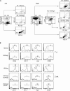
Phenotypic characterization of HSAhigh NKT cells. Thymocytes were MACS-enriched for tet+ cells and (A) analyzed for size (forward scatter, FSC) and granularity (side scatter, SSC) or (B) stained for HSA, CD44, NK1.1, Vβ8, or CD69. Line graphs display the frequency of CD44+, NK1.1+, Vβ8+, and CD69+ cells among tet+HSAlow or tet+HSAhigh cells gated as indicated. Data are representative of three individual experiments using pools of 7-d-old or 2-wk-old mice as indicated.
Figure 5.
Figure 5.
DN NKT cells arise from CD4+ precursors. NKT thymocyte subsets were isolated and FACS-sorted from 2–4-wk-old mice for intrathymic injection into NKT-deficient Jα18−/− recipients. The subsets injected included (A) tet+NK1.1CD4+, tet+NK1.1+CD4+, and tet+NK1.1+CD4 (as gated) or (B) CD4+CD44highNK1.1 cells (as gated) obtained from a preparation of HSAlowCD8-negative IAβb−/− thymocytes without tetramer staining. CD4/NK1.1 profiles of sorted cells and their progeny 7 d after intrathymic injection are shown, as indicated. Numbers represent the percentages of each subpopulation in indicated gate or quadrant. Data are representative of three separate experiments.
Figure 6.
Figure 6.
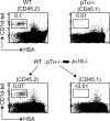
Cell-autonomous requirement for pTα in NKT thymocyte development. Mature NKT cells in WT, pTα−/−, and pTα−/−:WT bone marrow → Jα18−/− radiation chimeras. In the mixed chimera, the WT compartment is identified by CD45.2 gating and the pTα−/− compartment by CD45.1 as indicated. Data are representative of at least four mice in each group.
Figure 7.
Figure 7.
New model outlining the cellular developmental stages leading to mature CD4 and DN NKT cells. The dashed arrows indicate hypothetical precursor–product relationship.

References

    1. Park, S.H., and A. Bendelac. 2000. CD1-restricted T-cell responses and microbial infection. Nature. 406:788–792. - PubMed (VSports在线直播)
    1. Godfrey, D.I., K.J. Hammond, L.D. Poulton, M.J. Smyth, and A.G. Baxter. 2000. NKT cells: facts, functions and fallacies. Immunol. Today. 21:573–583. - PubMed (V体育安卓版)
    1. Kinjo, Y., D. Wu, G. Kim, G.W. Xing, M.A. Poles, D.D. Ho, M. Tsuji, K. Kawahara, C.H. Wong, and M. Kronenberg. 2005. Recognition of bacterial glycosphingolipids by natural killer T cells. Nature. 434:520–525. - PubMed
    1. Zhou, D., J. Mattner, C. Cantu III, N. Schrantz, N. Yin, Y. Gao, Y. Sagiv, K. Hudspeth, Y.P. Wu, T. Yamashita, et al. 2004. Lysosomal glycosphingolipid recognition by NKT cells. Science. 306:1786–1789. - "V体育安卓版" PubMed
    1. Mattner, J., K.L. DeBord, N. Ismail, R.D. Goff, C. Cantu III, D. Zhou, P. Saint-Mezard, V. Wang, Y. Gao, N. Yin, et al. 2005. Both exogenous and endogenous glycolipid antigens activate NKT cells during microbial infections. Nature. 434:525–529. - PubMed

Publication types

MeSH terms

Substances