Skip to main page content
U.S. flag

An official website of the United States government

Dot gov

The . gov means it’s official. Federal government websites often end in . gov or . mil. Before sharing sensitive information, make sure you’re on a federal government site VSports app下载. .

Https

The site is secure. The https:// ensures that you are connecting to the official website and that any information you provide is encrypted and transmitted securely. V体育官网.

Clinical Trial
. 2005 May 2;201(9):1503-17.
doi: 10.1084/jem.20042592.

Sustained expansion of NKT cells and antigen-specific T cells after injection of alpha-galactosyl-ceramide loaded mature dendritic cells in cancer patients

Affiliations
Clinical Trial

Sustained expansion of NKT cells and antigen-specific T cells after injection of alpha-galactosyl-ceramide loaded mature dendritic cells in cancer patients

David H Chang et al. J Exp Med. .

Erratum in

  • J Exp Med. 2007 Oct;204(10):2487

Abstract

Natural killer T (NKT) cells are distinct glycolipid reactive innate lymphocytes that are implicated in the resistance to pathogens and tumors. Earlier attempts to mobilize NKT cells, specifically, in vivo in humans met with limited success. Here, we evaluated intravenous injection of monocyte-derived mature DCs that were loaded with a synthetic NKT cell ligand, alpha-galactosyl-ceramide (alpha-GalCer; KRN-7000) in five patients who had advanced cancer. Injection of alpha-GalCer-pulsed, but not unpulsed, dendritic cells (DCs) led to >100-fold expansion of several subsets of NKT cells in all patients; these could be detected for up to 6 mo after vaccination. NKT activation was associated with an increase in serum levels of interleukin-12 p40 and IFN-gamma inducible protein-10 VSports手机版. In addition, there was an increase in memory CD8+ T cells specific for cytomegalovirus in vivo in response to alpha-GalCer-loaded DCs, but not unpulsed DCs. These data demonstrate the feasibility of sustained expansion of NKT cells in vivo in humans, including patients who have advanced cancer, and suggest that NKT activation might help to boost adaptive T cell immunity in vivo. .

PubMed Disclaimer (VSports)

Figures

Figure 1.
Figure 1.
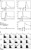
Expansion of NKT cells after injection of GalCer-pulsed DCs. (A) Kinetics of Vα24+/Vβ11+ NKT cells in blood. The proportion of Vα24+/Vβ11+ NKT cells in blood was monitored before and after DC vaccination. The dates of DC injections are depicted by arrows. (B) Representative FACS plots showing the number of invariant NKT cells before and after DC vaccination. Numbers in upper right quadrant represent percentage of NKT/total lymphocytes. (C) Correlation between kinetics of α-GalCer dimer binding cells and invariant TCR+ cells. Dimer positive NKT cells were analyzed based on the coexpression of Vα24 or Vβ11. The data shown are from K3. (D) Representative FACS plots showing the expression of Vα24/Vβ11+ on α-GalCer–CD1d dimer binding cells elicited after vaccination in a patient (K3). A sub-population of Vα24-CD1d dimer+ cells is circled. Note that the Vβ11+ dimer+ populations consist of two distinct subsets. (E) Expansion of invariant NKT cells in the tumor bed. NKT cells in the blood or marrow tumor bed were monitored by flow cytometry.
Figure 1.
Figure 1.
Expansion of NKT cells after injection of GalCer-pulsed DCs. (A) Kinetics of Vα24+/Vβ11+ NKT cells in blood. The proportion of Vα24+/Vβ11+ NKT cells in blood was monitored before and after DC vaccination. The dates of DC injections are depicted by arrows. (B) Representative FACS plots showing the number of invariant NKT cells before and after DC vaccination. Numbers in upper right quadrant represent percentage of NKT/total lymphocytes. (C) Correlation between kinetics of α-GalCer dimer binding cells and invariant TCR+ cells. Dimer positive NKT cells were analyzed based on the coexpression of Vα24 or Vβ11. The data shown are from K3. (D) Representative FACS plots showing the expression of Vα24/Vβ11+ on α-GalCer–CD1d dimer binding cells elicited after vaccination in a patient (K3). A sub-population of Vα24-CD1d dimer+ cells is circled. Note that the Vβ11+ dimer+ populations consist of two distinct subsets. (E) Expansion of invariant NKT cells in the tumor bed. NKT cells in the blood or marrow tumor bed were monitored by flow cytometry.
Figure 2.
Figure 2.
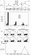
Kinetics of NKT subsets after the injection of α-GalCer–pulsed DCs. (A) Proportion of CD4+, double negative (DN) or CD8+ subsets of NKT cells after α-GalCer/DC vaccination in two patients (K2 and K3), as analyzed by flow cytometry. (B) Representative FACS plots from a patient (K2), showing subsets of mobilized NKT cells, at early, mid or later time points, as shown in (A). In the lower panel, numbers in the upper right quadrant are percentage of CD4+/double negative/CD8+, respectively of total NKT cells.
Figure 3.
Figure 3.
Cytokine multiplex analysis. Increase or decrease in serum levels before and 24 h after unpulsed (first DC injection) or α-GalCer–pulsed DC vaccine (third DC injection) were analyzed separately for each patient by Luminex. (A) Changes in serum IL-12p40. (B) Changes in serum MIP-1β. (C) Changes in serum IP-10.
Figure 4.
Figure 4.
Function of freshly isolated NKT cells mobilized in vivo. (A) Intracellular cytokine staining for α-GalCer reactive IFN-γ production. Freshly isolated PBMCs from postvaccination samples (K2–K5) containing NKT cells or healthy donors (H1–H5) were cultured overnight with anti-CD28 alone (as control [Ctrl]), or with 100 ng/ml α-GalCer (αGC) in the presence of monensin. The percentage of IFN-γ–producing Vα24+Vβ11+ cells was quantified by flow cytometry. The data shown are gated on Vα24+Vβ11+ cells. (B) Detection of α-GalCer reactive IFN-γ–producing cells by Elispot. Freshly isolated PBMCs from pre- or postvaccination samples were cultured overnight with 100 ng/ml α-GalCer. The presence of IFN-γ–producing cells was quantified by ELISPOT.
Figure 5.
Figure 5.
Proliferative capacity and function of mobilized NKT cells. (A) DC-mediated expansion of mobilized NKT cells in vitro. Pre- and postimmunization PBMCs were thawed together and cultured with autologous monocyte-derived mature DCs with or without pulsing with 100 ng/ml α-GalCer ([αGC]; DC/responder ratio of 1:20), in the presence of 50 U/ml IL-2. After 7 d of culture, the presence of Vα24+/Vβ11+ invariant NKT cells was quantified by flow cytometry. Numbers in the quadrant indicates the percentage of cells in the quadrant of the total lymphocyte population. (B) Binding of iNKT cells expanded in vitro from postvaccine samples to CD1d–α-GalCer dimers. Dimer(−) refers to unloaded dimers. Expression of Vα24 or Vβ11 on dimer-binding cells was analyzed by flow cytometry. (C) IFN-γ production by ex vivo expanded NKT cells. NKT cells expanded ex vivo as in (A) were stimulated overnight with unpulsed or α-GalCer–pulsed autologous DCs in the presence of monensin. NKT cells expanded from healthy donors (H6–H8) were used as a control. The presence of IFN-γ–producing cells was quantified by flow cytometry. (D) TaqMan analysis of cytokine secretion in expanded NKT cells. NKT cells expanded ex vivo as in (A) were stimulated overnight with unpulsed or α-GalCer–pulsed autologous DCs. NKT cells expanded from healthy donors (H9–H11) were used as a control. The presence of mRNA for selected cytokines (IFN-γ, IL-4, IL-10, and IL-13) was analyzed by real time RT-PCR. Data were normalized to the expression of a housekeeping gene, GAPDH. α-GalCer reactive transcription was analyzed by comparing the expression in control versus α-GalCer–stimulated samples. *P < 0.05 for comparison with healthy donor control.
Figure 5.
Figure 5.
Proliferative capacity and function of mobilized NKT cells. (A) DC-mediated expansion of mobilized NKT cells in vitro. Pre- and postimmunization PBMCs were thawed together and cultured with autologous monocyte-derived mature DCs with or without pulsing with 100 ng/ml α-GalCer ([αGC]; DC/responder ratio of 1:20), in the presence of 50 U/ml IL-2. After 7 d of culture, the presence of Vα24+/Vβ11+ invariant NKT cells was quantified by flow cytometry. Numbers in the quadrant indicates the percentage of cells in the quadrant of the total lymphocyte population. (B) Binding of iNKT cells expanded in vitro from postvaccine samples to CD1d–α-GalCer dimers. Dimer(−) refers to unloaded dimers. Expression of Vα24 or Vβ11 on dimer-binding cells was analyzed by flow cytometry. (C) IFN-γ production by ex vivo expanded NKT cells. NKT cells expanded ex vivo as in (A) were stimulated overnight with unpulsed or α-GalCer–pulsed autologous DCs in the presence of monensin. NKT cells expanded from healthy donors (H6–H8) were used as a control. The presence of IFN-γ–producing cells was quantified by flow cytometry. (D) TaqMan analysis of cytokine secretion in expanded NKT cells. NKT cells expanded ex vivo as in (A) were stimulated overnight with unpulsed or α-GalCer–pulsed autologous DCs. NKT cells expanded from healthy donors (H9–H11) were used as a control. The presence of mRNA for selected cytokines (IFN-γ, IL-4, IL-10, and IL-13) was analyzed by real time RT-PCR. Data were normalized to the expression of a housekeeping gene, GAPDH. α-GalCer reactive transcription was analyzed by comparing the expression in control versus α-GalCer–stimulated samples. *P < 0.05 for comparison with healthy donor control.
Figure 6.
Figure 6.
Changes in antigen-specific T cell responses. (A) Changes in CMVpp65 and Flu-MP–specific IFN-γ–producing T cells. Freshly isolated PBMCs were cultured overnight with defined A2-restricted peptides from Flu-MP or CMVpp65. The number of antigen-specific IFN-γ–producing T cells was measured using an Elispot assay. (B) Expansion of virus-specific memory T cells (recall tetramer assay). Samples from pre- or postimmunization samples were thawed together and cocultured with autologous mature DCs that were pulsed with specific peptides. After 7 d of culture, the number of peptide-specific MHC tetramer binding T cells was quantified by flow cytometry. (C) Representative FACS plots showing staining for CMVpp65-HLA A2 tetramer and Flu-MP–A2 tetramer, from an expansion as in (B). Number indicates percent of tetramer positive cells in the quadrant. (D) Changes in Flu-MP–specific memory T cells in a patient who also received an inactivated influenza vaccine during the course of the trial. Pre- and postimmunization T cells were thawed together and expanded with autologous peptide-pulsed DCs as in (B). After 7 d of culture, the presence of Flu-MP–A2 tetramer binding T cells was monitored by flow cytometry. SFC, spot-forming cells.
Figure 7.
Figure 7.
Changes in NK cells. The number of CD3CD56+ NK cells in fresh PBMCs was monitored by flow cytometry, before and after DC vaccination.
Figure 8.
Figure 8.
Schema for clinical trial.

"VSports最新版本" Comment in

  • Research on human subjects in the JEM.
    Steinman RM. Steinman RM. J Exp Med. 2005 May 2;201(9):1349-50. doi: 10.1084/jem.20050723. J Exp Med. 2005. PMID: 15867088 Free PMC article. No abstract available.

References

    1. Godfrey, D.I., and M. Kronenberg. 2004. Going both ways: immune regulation via CD1d-dependent NKT cells. J. Clin. Invest. 114:1379–1388. - "V体育ios版" PMC - PubMed
    1. Brigl, M., and M.B. Brenner. 2004. CD1: antigen presentation and T cell function. Annu. Rev. Immunol. 22:817–890. - PubMed
    1. Kawano, T., J. Cui, Y. Koezuka, I. Toura, Y. Kaneko, K. Motoki, H. Ueno, R. Nakagawa, H. Sato, E. Kondo, et al. 1997. CD1d-restricted and TCR-mediated activation of valpha14 NKT cells by glycosylceramides. Science. 278:1626–1629. - PubMed
    1. Stetson, D.B., M. Mohrs, R.L. Reinhardt, J.L. Baron, Z.E. Wang, L. Gapin, M. Kronenberg, and R.M. Locksley. 2003. Constitutive cytokine mRNAs mark natural killer (NK) and NK T cells poised for rapid effector function. J. Exp. Med. 198:1069–1076. - V体育安卓版 - PMC - PubMed
    1. Eberl, G., and H.R. MacDonald. 2000. Selective induction of NK cell proliferation and cytotoxicity by activated NKT cells. Eur. J. Immunol. 30:985–992. - PubMed

MeSH terms