<small lang="99GBw"></small> Skip to main page content
U.S. flag

An official website of the United States government

Dot gov

The . gov means it’s official. Federal government websites often end in . gov or VSports app下载. mil. Before sharing sensitive information, make sure you’re on a federal government site. .

Https

The site is secure V体育官网. The https:// ensures that you are connecting to the official website and that any information you provide is encrypted and transmitted securely. .

. 2005 May-Jun;28(3):258-67.
doi: 10.1097/01.cji.0000158855.92792.7a.

"V体育平台登录" Survival, persistence, and progressive differentiation of adoptively transferred tumor-reactive T cells associated with tumor regression

Affiliations

"V体育官网入口" Survival, persistence, and progressive differentiation of adoptively transferred tumor-reactive T cells associated with tumor regression

Jianping Huang et al. J Immunother. 2005 May-Jun.

Abstract

Objective clinical responses have been observed in approximately 50% of patients who received non-myeloablative chemotherapy prior to the adoptive transfer of autologous melanoma-reactive tumor-infiltrating lymphocytes (TILs). Recent studies carried out through the use of antibodies directed against T-cell-receptor beta chain variable region (TRBV) products, as well as by direct sequencing of the expressed TRBV gene products, indicated that clinical responses in this trial were associated with the level of persistence of adoptively transferred T cells VSports手机版. In an attempt to further characterize T cells that persist in vivo following adoptive transfer, five dominant T-cell clonotypes were identified in TIL 2035, an adoptively transferred TIL that was associated with the complete regression of multiple metastases. The most highly persistent clonotype, which expressed the BV1 TR gene product, recognized the MAGE-6 cancer/testis antigen in the context of HLA-A23. This clonotype was detected in peripheral blood for over 16 months following adoptive transfer, expressed relatively higher levels of the co-stimulatory markers CD28 and CD27, and possessed telomeres that were long relative to other clonotypes present in TIL 2035 that showed only short-term persistence. The long-term persistent BV1 clonotype appeared to differentiate more slowly toward an end-stage effector in vivo than short-term persistent clonotypes, as manifested by the downregulation of CD28, CD27, and CD45RO and upregulation of CD57 and CD45RA expression on these T cells. These results indicated that the differentiation stage and replicative history of individual TIL clonotypes might be associated with their ability to survive and to persist in vivo, and progressive differentiation of the persistent clonotypes occurred following adoptive transfer. .


Figures

FIGURE 1
FIGURE 1
Characterization of the antigen reactivity of TIL 2035 T cells. A, TIL 2035 cells were co-cultured with either autologous melanoma line 2035mel (HLA-A2, A23) or HLA-mismatched control melanoma line 888mel (HLA-A1, A24) for 6 hours, followed by incubation with an anti-CD107a monoclonal antibody and FACS analysis. Stimulations carried out with 2035 mel are indicated by the bold lines, and those carried out with control 888 mel are indicated by the filled histograms. Gating was carried out on the cell populations that expressed the indicated BV gene products. B, The autologous melanoma line and HLA-A2-positive melanoma cell line, 624 mel, were incubated with either no antibody (Control), anti-HLA-A*02 antibody (Anti-A2) or anti-HLA-A23 antibody (Anti-A23). TIL 2035-F6/8 was then added, and 18 hours later IFNγ release was measured by ELISA. C, Autologous EBVB cells were pulsed with either a control peptide (upper left) or the HLA-A2-restricted NY-ESO-1: 157-165 peptide (upper right) for 2 hours, and then co-cultured with TIL 2035-F6/8 for 2 hours. An IFNγ capture assay was then carried out, cells were co-stained with an anti-BV17 antibody, and FACS analysis was performed. Two hundred ninety-three cells were co-transfected HLA-A23 with either a control vector (lower left) or a vector encoding MAGE-6 (lower right) for 24 hours, followed by the addition of TIL 2035-F6/8. Eighteen hours later, an IFNγ capture assay was carried out; cells were co-stained with an anti-BV1 antibody and analyzed using FACS analysis. The data represent one of three individual experiments.
FIGURE 2
FIGURE 2
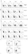
Phenotypic analysis of dominant T-cell populations in the infusion TIL 2035 and in peripheral blood. The infusion TIL 2035 as well as peripheral blood samples obtained 8 days or 20 days following adoptive transfer were gated on CD8+ cells and analyzed for expression of CD28 (A) and CD27 (B). The MFI of CD28 expression for the gated populations was 29 (BV1), 9 (BV2), 12 (BV14), and 6 (BV17). C, Co-expression of CD27 and CD28 was analyzed on TIL 2035 populations that were gated using the indicated TRBV antibodies. Quadrants were drawn based on staining obtained with IgG control antibodies. The data represent one of three individual experiments.
FIGURE 3
FIGURE 3
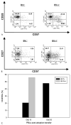
Expression of differentiation markers on PBMCs obtained following adoptive transfer. Co-expression of CD28 and CD57 (A) and CD27 and CD57 (B) was analyzed on peripheral blood samples obtained 12 days after adoptive transfer that were gated using the indicated BV antibody. Quadrants were drawn based on staining obtained with IgG control antibodies. C, Expression of CD45RA was evaluated for populations of PBMCs obtained 13 or 61 days following transfer that were gated using the indicated BV antibodies. Data represent one of three individual experiments.
FIGURE 4
FIGURE 4
Telomere fluorescence intensity of dominant clonotypes present in TIL 2035. BV1+ and BV14+ T cells were isolated from TIL 2035, and FISH was carried out using a telomeric peptide nucleic acid (PNA) probe, or a control PNA probe for each of the separated populations, followed by FACS analysis. The control staining corresponds to that observed for BV1+ T cells. Data represent one of two individual experiments.
FIGURE 5
FIGURE 5
Alterations in the phenotype BV1+ T cells in peripheral blood following adoptive transfer. Gated populations of BV1+ T cells present in peripheral blood samples 13 days, 61 days, 280 days, and 480 days after the adoptive transfer were analyzed for expression of (A) CD27, (B) CD28, (C) CD45RO, or (D) CD57. The day 0 sample represents TIL 2035. Data represent one of three individual experiments.

References

    1. Dudley ME, Wunderlich JR, Robbins PF, et al. Cancer regression and autoimmunity in patients after clonal repopulation with antitumor lymphocytes. Science. 2002;298:850–854. - PMC - PubMed
    1. Robbins PF, Dudley ME, Wunderlich JR, et al. Persistence of transferred lymphocyte clonotypes correlates with cancer regression in patients receiving cell transfer therapy. J Immunol. 2004;173:7125–7130. - VSports - PMC - PubMed
    1. Rufer N, Zippelius A, Batard P, et al. Ex vivo characterization of human CD8+ T subsets with distinct replicative history and partial effector functions. Blood. 2003;102:1779–1787. - PubMed (VSports app下载)
    1. Hendriks J, Xiao Y, Borst J. CD27 promotes survival of activated T cells and complements CD28 in generation and establishment of the effector T cell pool. J Exp Med. 2003;198:1369–1380. - PMC - PubMed
    1. Jones RG, Elford AR, Parsons MJ, et al. CD28-dependent activation of protein kinase B/Akt blocks Fas-mediated apoptosis by preventing death-inducing signaling complex assembly. J Exp Med. 2002;196:335–348. - "VSports手机版" PMC - PubMed

MeSH terms

LinkOut - more resources (VSports注册入口)