Skip to main page content
U.S. flag

An official website of the United States government

Dot gov

The . gov means it’s official. Federal government websites often end in . gov or . mil VSports app下载. Before sharing sensitive information, make sure you’re on a federal government site. .

Https

The site is secure V体育官网. The https:// ensures that you are connecting to the official website and that any information you provide is encrypted and transmitted securely. .

. 2005 Mar 24:6:71.
doi: 10.1186/1471-2105-6-71.

Prediction of a common structural scaffold for proteasome lid, COP9-signalosome and eIF3 complexes (VSports注册入口)

Affiliations

Prediction of a common structural scaffold for proteasome lid, COP9-signalosome and eIF3 complexes

Hartmut Scheel et al. BMC Bioinformatics. .

Abstract

Background: The 'lid' subcomplex of the 26S proteasome and the COP9 signalosome (CSN complex) share a common architecture consisting of six subunits harbouring a so-called PCI domain (proteasome, CSN, eIF3) at their C-terminus, plus two subunits containing MPN domains (Mpr1/Pad1 N-terminal) VSports手机版. The translation initiation complex eIF3 also contains PCI- and MPN-domain proteins, but seems to deviate from the 6+2 stoichiometry. Initially, the PCI domain was defined as the region of detectable sequence similarity between the components mentioned above. .

Results: During an exhaustive bioinformatical analysis of proteasome components, we detected multiple instances of tetratrico-peptide repeats (TPR) in the N-terminal region of most PCI proteins, suggesting that their homology is not restricted to the PCI domain. We also detected a previously unrecognized PCI domain in the eIF3 component eIF3k, a protein whose 3D-structure has been determined recently V体育安卓版. By using profile-guided alignment techniques, we show that the structural elements found in eIF3k are most likely conserved in all PCI proteins, resulting in a structural model for the canonical PCI domain. .

Conclusion: Our model predicts that the homology domain PCI is not a true domain in the structural sense but rather consists of two subdomains: a C-terminal 'winged helix' domain with a key role in PCI:PCI interaction, preceded by a helical repeat region. The TPR-like repeats detected in the N-terminal region of PCI proteins most likely form an uninterrupted extension of the repeats found within the PCI domain boundaries. This model allows an interpretation of several puzzling experimental results V体育ios版. .


Figures

Figure 1
Figure 1
TPR-like motifs upstream and inside the PCI domain. The proposed domain topologies of selected human PCI proteins were investigated by profile techniques. Besides the common PCI domain (blue) short stretches of ~35 aa each are depicted in orange and light blue. These stretches show weak to medium similarity to TPR segments in established TPR proteins and merge seamlessly into the PCI domain in several PCI subunits. Corresponding accession numbers are listed in figure 2.
Figure 2
Figure 2
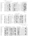
Multiple sequence alignment of human PCI subunits from proteasome lid, CSN and eIF3. Shown are only the segments matched by the PROSITE PCI domain. Conserved residues printed on black background were found in at last 50 % of ~60 PCI proteins from selected species, from which only human representatives are shown. Grey background was assigned to positions occupied by residues with similar physicochemical properties in at least 50 % of the sequences. The alignment was shaded using BOXSHADE . Above the PCI alignment secondary structure prediction as calculated from JPred [31] is presented. In these calculations sequences of eIF3k homologues were not included. Secondary structure elements of eIF3k as derived from PDB structure 1RZ4 are shown in a separate row. The abbreviations denote the following secondary structure types: E extended (sheet) and H helix. In addition, structural subdomain classification ('HAM', 'WH') as described in Wei et al. [26] and domain boundaries according to PCI profiles from PROSITE and Pfam are provided. Sequence names correspond to the following SwissProt database entries: eIF3k (Q9UBQ5), PSMD3 (O43242), PSMD6 (Q15008), PSMD8 (P48556), PSMD11 (O00231), PSMD12 (O00232), PSMD13 (Q9UNM6), CSN1 (Q13098), CSN2 (P61201), CSN3 (Q9UNS2), CSN4 (Q9BT78), CSN8 (Q99627).
Figure 3
Figure 3
(A) shows the overall structure of eIF3k from the PDB-entry 1RZ4 [26] with β-strands and α-helices represented as ribbons and cylinders, respectively. Regions of the structure with sequence similarity to canonical PCI domain are rendered in colour. Regions belonging to the WH subdomain are shown in green, while conserved structure elements of the helical hairpin regions are shown in dark blue. The connection between β-strand 2 and 3 is not resolved and thus missing in 1RZ4. Other regions (extreme N- and C- termini, connecting helices between hairpins, unstructured regions) are shown in grey. (B) Model of a PCI protein with three additional helical hairpins upstream of the PCI domain. Within the PCI domains, only regions that can be modelled on the eIF3k template are shown. The N-terminal extension is shown in light blue, the other colours are as in figure 3a.

References

    1. Kim T, Hofmann K, von Arnim AG, Chamovitz DA. PCI complexes: pretty complex interactions in diverse signaling pathways. Trends Plant Sci. 2001;6:379–386. doi: 10.1016/S1360-1385(01)02015-5. - DOI - PubMed
    1. Hofmann K, Bucher P. The PCI domain: a common theme in three multiprotein complexes. Trends Biochem Sci. 1998;23:204–205. doi: 10.1016/S0968-0004(98)01217-1. - VSports注册入口 - DOI - PubMed
    1. Aravind L, Ponting CP. Homologues of 26S proteasome subunits are regulators of transcription and translation. Protein Sci. 1998;7:1250–1254. - VSports在线直播 - PMC - PubMed
    1. Kapelari B, Bech-Otschir D, Hegerl R, Schade R, Dumdey R, Dubiel W. Electron microscopy and subunit-subunit interaction studies reveal a first architecture of COP9 signalosome. J Mol Biol. 2000;300:1169–1178. doi: 10.1006/jmbi.2000.3912. - DOI - PubMed
    1. Fu H, Reis N, Lee Y, Glickman MH, Vierstra RD. Subunit interaction maps for the regulatory particle of the 26S proteasome and the COP9 signalosome. Embo J. 2001;20:7096–7107. doi: 10.1093/emboj/20.24.7096. - "VSports app下载" DOI - PMC - PubMed

MeSH terms

LinkOut - more resources