Skip to main page content
U.S. flag

An official website of the United States government

Dot gov

The VSports app下载. gov means it’s official. Federal government websites often end in . gov or . mil. Before sharing sensitive information, make sure you’re on a federal government site. .

Https

The site is secure. The https:// ensures that you are connecting to the official website and that any information you provide is encrypted and transmitted securely. V体育官网.

Review
. 2004 Dec;68(4):692-744.
doi: 10.1128/MMBR.68.4.692-744.2004.

Type V protein secretion pathway: the autotransporter story

Affiliations
Review

Type V protein secretion pathway: the autotransporter story (VSports手机版)

"VSports在线直播" Ian R Henderson et al. Microbiol Mol Biol Rev. 2004 Dec.

Abstract

Gram-negative bacteria possess an outer membrane layer which constrains uptake and secretion of solutes and polypeptides. To overcome this barrier, bacteria have developed several systems for protein secretion. The type V secretion pathway encompasses the autotransporter proteins, the two-partner secretion system, and the recently described type Vc or AT-2 family of proteins. Since its discovery in the late 1980s, this family of secreted proteins has expanded continuously, due largely to the advent of the genomic age, to become the largest group of secreted proteins in gram-negative bacteria. Several of these proteins play essential roles in the pathogenesis of bacterial infections and have been characterized in detail, demonstrating a diverse array of function including the ability to condense host cell actin and to modulate apoptosis. However, most of the autotransporter proteins remain to be characterized VSports手机版. In light of new discoveries and controversies in this research field, this review considers the autotransporter secretion process in the context of the more general field of bacterial protein translocation and exoprotein function. .


Figures

FIG. 1.
FIG. 1.
Schematic representation of the type I, II, III, and IV protein secretion systems. The type I pathway is exemplified by hemolysin A (HlyA) secretion in E. coli, the type III system is exemplified by Yop secretion in Yersinia, the type II system is exemplified by pullulanase secretion in Klebsiella oxytoca, and the type IV system is exemplified by the VirB system in A. tumefaciens. ATP hydrolysis by HlyB, YscN, SecA, and VirB11 is indicated. Secreted effector molecules are depicted as grey ovals. The type II and in some cases the type IV secretion systems utilize the cytoplasmic chaperone SecB, although the Tat export pathway has recently been implicated in the secretion of molecules via the type II pathway. Type III secretion also involves cytoplasmic chaperones (SycE); however, they do not interact with the Sec inner membrane translocon. The major structural proteins of each system are depicted in relation to their known or deduced position in the cell envelope. EM, extracellular milieu; OM, outer membrane; Peri, periplasm; IM, inner membrane; Cyto, cytoplasm.
FIG. 2.
FIG. 2.
Structure of TolC. The structure of TolC was solved to 2.1-Å resolution. The functional TolC unit consists of three TolC monomers oligomerized into a regular structure embedded in the outer membrane and extending into the periplasm such that it forms a continuous solvent-accessible conduit. The portion of the trimeric molecule that is embedded in the outer membrane adopts a 12-strand β-barrel conformation consisting of four β-strands per monomer. The molecule is 140 Å long, the β-barrel is 40 Å long (spanning the outer membrane), and the remaining portion of the molecule adopts a 100 Å α-helical conformation spanning the periplasmic space. This α-helical domain consists of 12 α-helices, such that the α-helices extend from or connect to the periplasmic side of each β-strand. It is the α-helical domain that is predicted to interact with the MFP. Each monomer is highlighted in a separate color. Adapted from reference and the Protein Data Bank (30).
FIG. 3.
FIG. 3.
Schematic overview of the type V secretion systems. The secretion pathway of the autotransporter proteins (type Va) is depicted at the bottom left of the diagram, the two-partner system (type Vb) is depicted in the center of the diagram, and the type Vc or AT-2 family is depicted on the right. The four functional domains of the proteins are shown: the signal sequence, the passenger domain, the linker region, and the β-domain. The autotransporter polyproteins are synthesized and generally exported through the cytoplasmic membrane via the Sec machinery. Interestingly, effector proteins with an unusual extended signal sequence, which purportedly mediates Srp-dependent export, are found in all three categories of type V secretion. Once through the inner membrane, the signal sequence is cleaved and the β-domain inserts into the outer membrane in a biophysically favored β-barrel structure that forms a pore in the outer membrane. After formation of the β-barrel, the passenger domain inserts into the pore and is translocated to the bacterial cell surface, where it may or may not undergo further processing.
FIG. 4.
FIG. 4.
Structure and alignment of the extended signal sequences. The extended signal sequences belonging to autotransporter proteins from a wide range of gram-negative bacteria are depicted. Blue shading indicates the positions of the positively charged n1 and n2 domains. The positions of the hydrophobic h1 and h2 domains are denoted by yellow shading. The signal peptidase recognition sites are indicated by green shading. The n2, h2 and C-domains are characteristic of a typical signal sequence secreted via the Sec-dependent translocon in conjunction with the SecB chaperone. The in silico-predicted and/or empirically determined cleavage site between the signal sequence and the passenger domain is indicated. Conserved residues are highlighted.
FIG. 5.
FIG. 5.
Alternative mechanisms of autotransporter protein secretion. (A) Several different hypotheses have been proffered for the mechanism of autotransporter protein biogenesis. From left to right are depicted the traditional view of secretion originally proposed by Pohlner et al. (401), in which the passenger domain passes through the pore of the β-barrel to the outside; a recently proposed structure where the passenger domain extends directly from the β-barrel into the extracellular milieu; the secretion pathway proposed by Veiga et al. (512), in which a central channel is formed through which the passenger domains are secreted to the outside, and the pathway proposed by Hoiczyk et al. (210) for the type Vc autotransporter family, in which several molecules contribute β-strands to make a large β-barrel pore through which the proteins are secreted to the external side of the outer membrane. (B) Crystal structure of the AspA/NalP translocating unit to 2.6 Å. A side view and a stereo-top view are depicted. The protein forms a 12-strand β-barrel structure characterized by short periplasmic turns and longer external loops. The barrel interior is highly hydrophilic due to the presence of charged amino acids. Within the barrel is embedded an α-helical region, which is attached to the first transmembrane β-strand such that the extreme N terminus of the protein, and to which it is presumed a native passenger domain would be attached, is located on the extracellular surface. Adapted from reference and the Protein Data Bank (30).
FIG. 6.
FIG. 6.
Phylogenetic tree of the autotransporter proteins. The CLUSTALX and TREE programs were used for multiple alignments and construction of a phylogenetic tree for the functionally characterized autotransporter proteins. The tree depicted in this figure is derived from analyses of the C-terminal translocating domains of the autotransporters. Proteins are clustered according to the pattern proposed by Yen et al. (540). A single additional cluster (cluster 11) is indicated.
FIG. 7.
FIG. 7.
Phase-variable expression of antigen 43. (A) Crossed-immunoelectrophoresis profile of Triton X-100-EDTA-extracted E. coli membrane vesicles. This represents the first resolution of the antigen 43 complex. (B) Coomassie blue-stained SDS-PAGE profile of outer membrane proteins from antigen 43-expressing and nonexpressing variants of E. coli. (C and D) Fluorescence microscopy of antigen 43 phase ON (Ag43+) and phase OFF (Ag43) populations of E. coli, respectively. (E) Expression of antigen 43 induces bacteria to autoaggregate and thus to settle to the bottom of the growth vessel. The identity of antigen 43, its α43 and β43 subunits, and expressing and nonexpressing variants are depicted by arrowheads and appropriate labels. (Panels A, B, and E, copyright Peter Owen and Mary Meehan.)
FIG. 8.
FIG. 8.
IcsA-mediated Shigella motility. Fluorescence microscopy demonstrating that IcsA (red label) is expressed preferentially at one pole of the bacterium, where it mediates actin (green label) polymerization and formation of the characteristic comet tails. The actin comet tails are indicated by white arrowheads, and the Shigella bacterium is indicated by a black arrowhead. (Copyright Marcia B. Goldberg.)
FIG. 9.
FIG. 9.
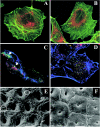
Activity of the EAEC Pet toxin. (A and B) Internalization of Pet (A) and a serine protease motif mutant (Pet S260I) (B) into HEp-2 cells. HEp-2 cells were treated with either Pet or Pet S260I for 1 h. The actin cytoskeleton is labeled with green, and Pet or Pet S2601 is labeledwith red. Note the perinuclear localization of Pet or Pet S260I inside the cells. (C and D) Effect of Pet (C) or Pet S260I (D) on fodrin redistribution in HEp-2 cells. HEp-2 cells were treated with either Pet or Pet S260I for 3 h. The actin cytoskeleton is labeled with blue, Pet or Pet S260I is labeled with red, and fodrin is labeled with green. Note that Pet but not Pet S260I causes cytoskeletal damage and fodrin redistribution (arrows), and it is possible to detect a delayed interaction between Pet S260I and fodrin due to inability to cleave it (yellow dots). (E and F) Scanning electron photomicrographs of in vitro-cultured human colonic tissues infected with the EAEC strain 042 (E) and the pet mutant strain J1F1 (F). For the tissue shown in panel E, the surface of the colon is markedly abnormal, as manifested by increased crypt apertures (white arrowhead), prominent mucosal crevices (white arrow), goblet cell pitting (black arrowhead in a circle), and rounding of epithelial cells (black arrow in a circle).
FIG. 10.
FIG. 10.
Phylogenetic relationships of the transporter domains of known and predicted B. bronchiseptica autotransporters. The predicted transporter domains of B. bronchiseptica autotransporters were aligned using CLUSTALW and subjected to distance matrix and neighbor-joining methods by using the PHYLIP package. The resulting phylogenetic tree was visualized with TreeView. Bootstrap values are for 100 replicates.
FIG. 11.
FIG. 11.
Crystal structure of the B. pertussis protein pertactin. The structure of pertactin was solved to a resolution of 2.5 Å (123). (A) Side view of pertactin, with the N terminus located to the top. (B) End view of pertactin looking from the N terminus toward the C terminus. This protein consists of 16 parallel β-strands arranged in a left-handed helix from which several loops extend. T1, T2, and T3 represent the turns between the three strands of the β-helix. The RGD motif is shown in purple in T1. The region in blue represents a conserved region that is present in many autotransporters and has been shown to mediate folding of the passenger domain (351). This is the largest β-helix known. Structurally, this molecule is related to the tail spike proteins of P22 phage and to the pectin lyases and methylesterases (227). Adapted from reference and the Protein Data Bank (30).

References

    1. Aguirre, P. M., J. B. Cacho, L. Folgueira, M. Lopez, J. Garcia, and A. C. Velasco. 1990. Rapid fluorescence method for screening Salmonella spp. from enteric differential agars. J. Clin. Microbiol. 28:148-149. - PMC - PubMed
    1. Ahl, T., and J. Reinholdt. 1991. Detection of immunoglobulin A1 protease-induced Fab alpha fragments on dental plaque bacteria. Infect. Immun. 59:563-539. - PMC - PubMed
    1. Ait-Tahar, K., K. G. Wooldridge, D. P. Turner, M. Atta, I. Todd, and D. A. Ala'Aldeen. 2000. Auto-transporter A protein of Neisseria meningitidis: a potent CD4+ T-cell and B-cell stimulating antigen detected by expression cloning. Mol. Microbiol. 37:1094-1105. - PubMed
    1. Aldridge, P., and K. T. Hughes. 2001. How and when are substrates selected for type III secretion? Trends Microbiol. 9:209-214. - VSports最新版本 - PubMed
    1. Al-Hasani, K., I. R. Henderson, H. Sakellaris, K. Rajakumar, T. Grant, J. P. Nataro, R. Robins-Browne, and B. Adler. 2000. The sigA gene which is borne on the she pathogenicity island of Shigella flexneri 2a encodes an exported cytopathic protease involved in intestinal fluid accumulation. Infect. Immun. 68:2457-2463. - PMC - PubMed

LinkOut - more resources