Skip to main page content
U.S. flag

An official website of the United States government

Dot gov

The VSports app下载. gov means it’s official. Federal government websites often end in . gov or . mil. Before sharing sensitive information, make sure you’re on a federal government site. .

Https

The site is secure V体育官网. The https:// ensures that you are connecting to the official website and that any information you provide is encrypted and transmitted securely. .

. 2004 Dec;78(23):13173-81.
doi: 10.1128/JVI.78.23.13173-13181.2004.

Human cytomegalovirus-specific CD4+-T-cell cytokine response induces fractalkine in endothelial cells

Affiliations

Human cytomegalovirus-specific CD4+-T-cell cytokine response induces fractalkine in endothelial cells (VSports注册入口)

V体育ios版 - Cynthia A Bolovan-Fritts et al. J Virol. 2004 Dec.

Abstract

Cytomegalovirus (CMV) infection has been linked to inflammation-related disease processes in the human host, including vascular diseases and chronic transplant rejection. The mechanisms through which CMV affects the pathogenesis of these diseases are for the most part unknown. To study the contributing role of the host immune response to CMV in these chronic inflammatory processes, we examined endothelial cell interactions with peripheral blood mononuclear cells (PBMC). Endothelial cultures were monitored for levels of fractalkine induction as a marker for initiating the host inflammatory response VSports手机版. Our results demonstrate that in the presence of CMV antigen PBMC from normal healthy CMV-seropositive donors produce soluble factors that induce fractalkine in endothelial cells. This was not observed in parallel assays with PBMC from seronegative donors. Examination of subset populations within the PBMC further revealed that CMV antigen-stimulated CD4(+) T cells were the source of the factors, gamma interferon and tumor necrosis factor alpha, driving fractalkine induction. Direct contact between CD4(+) cells and the endothelial monolayers is required for this fractalkine induction, where the endothelial cells appear to provide antigen presentation functions. These findings indicate that CMV may represent one member of a class of persistent pathogens where the antigen-specific T-cell response can result in the induction of fractalkine, leading to chronic inflammation and endothelial cell injury. .


Figures
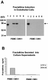
FIG. 1.
FIG. 1.
Fractalkine induction in endothelial cell cultures associated with CMV antigen-stimulated PBMC from CMV-seropositive donors. The data are representative of three separate experiments with different blood donors. (A) Detection of fractalkine induction in endothelial cell monolayers cocultured with CMV-seropositive PBMC by Western blot analysis with fractalkine-specific antibody. Lanes in left gel panel: 1, positive control for fractalkine, with ∼85-kDa form of fractalkine (note that the fractalkine form [R&D] lacks 57 carboxy-terminal amino acids and migrates at ∼85 kDa, whereas the endothelial-cell-associated form [also from R&D] is full length and migrates at ∼100 kDa); 2, negative control for fractalkine with endothelial cells in a resting state; 3 and 4, replicate samples of CMV antigen-treated PBMC from a CMV-seropositive donor; 5 and 6, replicate samples of media alone-treated PBMC from a CMV-seropositive donor. Lanes in right gel panel: 1, positive control for fractalkine, with ∼85-kDa form of fractalkine (commercially available from R&D); 2, negative control for fractalkine with endothelial cells in a resting state; 3 and 4, replicate samples of CMV antigen-treated PBMC from a CMV-seronegative donor; 5 and 6, replicate samples of medium-alone-treated PBMC from a CMV-seronegative donor. The arrow at the right of the gel indicates the Mr of fractalkine. (B) Detection of secreted fractalkine levels in coculture supernatants. ELISA results from coculture supernatants, comparing CMV-seropositive PBMC to CMV-seronegative PBMC.
FIG. 2.
FIG. 2.
Western blot analysis showing fractalkine induction is associated with CMV-seropositive lymphoid populations. The data are representative of two separate experiments with different blood donors. A CD14+ sample analysis is shown in the left panel, with lanes loaded as follows: lane 1, positive control for fractalkine with ∼85-kDa form of protein; lane 2, negative control for fractalkine with endothelial cells in a resting state; lanes 3 and 4, replicate samples of CMV antigen-treated cocultures with CD14+ and AEC; lanes 5 and 6, replicate cocultures of CD14+ cells and AEC in medium alone. A lymphoid-enriched fraction was used in a parallel analysis (right panel): lane 1, positive control for fractalkine with ∼85-kDa form of the protein (R&D); lane 2, negative control for fractalkine with endothelial cells in a resting state; lanes 3 and 4, replicate samples of CMV antigen-treated cocultures with lymphoid-enriched cells and AEC; lanes 5 and 6, replicate cocultures of lymphoid-enriched cells and AEC in medium alone.
FIG. 3.
FIG. 3.
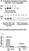
Fractalkine induction associated with CD4+ T-cell population. The data are representative of three experiments with different donors. (A) Western blots showing fractalkine induction in AEC cocultured with CD4+ cells, CD4+ CD8+ cells, and CD8+ cells, each stimulated with HCMV antigen and as nonstimulated resting state controls. The left panel shows results for CD4+ T cells with lanes loaded as follows: lane 1, positive control for fractalkine (R&D); lanes 2 and 3, replicate samples of CMV antigen-treated CD4+ T cells from a CMV-seropositive donor; lanes 4 and 5, replicate samples of medium-alone-treated same CD4+ T cells from a CMV-seropositive donor. The arrow between panels indicates the relative mobility of fractalkine. The right panel shows the results for CD4+ CD8+ samples, with lanes loaded as follows: lane 1, positive control for fractalkine; lanes 2 and 3, replicate samples of CMV antigen-treated CD4+ CD8+ T cells from a CMV-seropositive donor; lanes 4 and 5, replicate samples of medium-alone-treated CD4+ CD8+ cells; lanes 6 and 7, replicate samples of CMV antigen-treated CD8+ T cells; lanes 8 and 9, replicate samples of medium-alone-treated samples of CD8+ T cells from a CMV-seropositive donor. (B) ELISA demonstrating presence of secreted fractalkine in coculture medium with CD4+ cells in presence of CMV antigen and AEC. In contrast to this, CD8+ cells from CMV-seropositive donors show a clear absence of fractalkine production with CD8+ cells cocultured with AEC, even in the presence of CMV antigen.
FIG. 4.
FIG. 4.
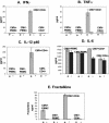
Screening for candidate factors (IFN-γ, TNF-α, IL-12, and IL-6) by ELISA to determine which factors are associated consistently with fractalkine induction by CMV antigen stimulation. A “+” or “−” symbol below the bar graphs refers to samples with or without CMV antigen stimulation, respectively. Supernatant samples were screened from day 3 cocultures of AEC monolayers with PBMC or CD4+ cells from a CMV-seropositive and CMV-seronegative pair of donors. (A) IFN-γ; (B) TNF-α; (C) IL-12; (D) IL-6; (E) fractalkine.
FIG. 5.
FIG. 5.
Reconstruction assays in which the candidate factors IFN-γ, TNF-α, and IL-12 were tested for their ability to directly induce fractalkine in endothelial cultures, with the levels of each factor as measured on day 3 in supernatants from cocultivations. The graph shows the levels of induced fractalkine resulting from each cytokine or combination as detected by ELISA screen for fractalkine secreted into supernatants. Specific cytokines and concentrations are represented on the x axis as follows: a, 200 pg of IL-12/ml; b, 400 pg of IL-12/ml; c, 500 pg of IFN-γ/ml; d, 50 pg of TNF-α/ml; e, 200 pg of IL-12/ml + 50 pg of TNF-α/ml; f, 200 pg of IL-12/ml + 50 pg of TNF-α/ml; g, 50 pg of TNF-α/ml + 500 pg of IFN-γ/ml; h, 200 pg of IL-12/ml + 500 pg of IFN-γ/ml + 50 pg of TNF-α/ml; i, CMV + CD4+ cells + CMV antigen-stimulated AEC coculture supernatants day 3; j, 50 ng of TNF-α/ml + 1,000 pg of IFN-γ/ml; k, medium alone.
FIG. 6.
FIG. 6.
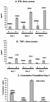
Time course in CMV antigen-stimulated CD4+ lymphocytes and PBMC cocultured with endothelial cells, showing cumulative levels of TNF-α (A) and IFN-γ (B) in culture supernatants over the course of 3 days. The results are shown with a CMV-seropositive donor, since the CMV-seronegative donor was completely negative for fractalkine induction. (C) Cumulative fractalkine levels by day 3 from same cocultures, showing results from a CMV-seropositive donor and a CMV-seronegative donor as a control. The data are representative of three separate experiments with different donors. The “+” or “−” symbol below graphs refer to samples with or without CMV antigen stimulation, respectively.
FIG. 7.
FIG. 7.
Neutralization assays confirm TNF-α and IFN-γ drive fractalkine induction in AEC. Neutralization carried out directly in cocultivations of CD4+ lymphocytes or PBMC with AEC. (A) Demonstration by ELISA screens of culture supernatants, showing loss of fractalkine in supernatants with neutralizing antibodies to target TNF-α and IFN-γ. In this sample, CD4+ cells were set up to scale with PBMC. Specific antibody blocks and controls are identified on the x axis as follows: No Ab, positive control with no antibody block; Iso, isotype control antibody; TNFa Ab block, antibody-treated sample to specifically neutralize TNF-α; IFNg Ab block, specific neutralization with IFN-γ antibody; Dual Ab block, combined anti-IFNγ and TNF-α antibodies; IL-12 Ab block, antibody to specifically neutralize IL-12. (B) In this sample, CD4+ cells are not set to scale but were loaded at ∼106 cells per well, ∼4-fold above scale to the level of CD4+ cells in PBMC. Antibody blocks and controls are as described for panel A on the x axis; “Dual Ab block per day” refers to combined anti-IFN-γ and TNF-α antibodies added on each of the 3 days during cocultivation. The data are representative of three separate experiments with different donor samples.
FIG. 8.
FIG. 8.
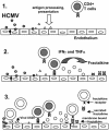
Proposed model for role of fractalkine induction by CMV antigen-specific CD4+ response leading to endothelial damage. CMV infection in endothelial cells releases viral proteins as shown in panel 1. These are processed and presented as antigen by other adjacent endothelial cells as shown in panel 2. CMV viral antigen is presented to CMV antigen-specific CD4+ cells by the endothelial cells. IFN-γ and TNF-α are released by the antigen-stimulated CD4+ cells (represented by cells with gray-shaded nuclei); these released factors interact with the endothelial cells, resulting in fractalkine induction (panel 3). Localized concentrations of fractalkine on the surface of endothelial cells, together with the release of soluble fractalkine, results in a fractalkine gradient. This can attract additional inflammatory cells to the site (these cells are represented by nonshaded nuclei), including monocyte/macrophage cells, NK cells, and CD8+ populations. Also, the presence of CMV US28, a CX3CR1 mimic, on the surface of endothelial cells during infection and on the envelope of virion particles released in the localized area could bind fractalkine and influence the inflammation processes dependent on fractalkine signals. This illustration is expanded from the fractalkine and vascular injury model of H. Umehara (30).

References

    1. Ancuta, P., R. Rao, A. Moses, A. Mehle, S. K. Shaw, F. W. Luscinskas, and D. Gabuzd. 2003. Fractalkine preferentially mediates arrest and migration of CD16+ monocytes. J. Exp. Med. 197:1701-1707. - PMC - PubMed
    1. Bettinotti, M. P., S. Solomon, M. Battiwala, N. Hensel, K. Ghazarian, D. Stroncek, and J. Barrett. 2003. T-cell response against the cytomegalovirus proteins pp65 and IE1. Hum. Immunol. 64:S73.
    1. Bitmanseur, A. D., S. L. Waldrop, C. J. Pitcher, E. Khatamzah, F. Kern, V. C. Maino, and L. J. Picker. 2001. Clonotypic structure of the human CD4+ memory T-cell response to CMV. J. Immunol. 167:1151-1163. - PubMed (VSports app下载)
    1. Bruning, J. H., M. C. Persoons, K. B. Lemstrom, F. S. Stals, E. De Clercq, and C. A. Bruggeman. 1994. Enhancement of transplantation-associated atherosclerosis by CMV, which can be prevented by antiviral therapy in the form of HPMPC. Transplant. Int. 7(Suppl. 1):S365-S370. - PubMed
    1. Compton, T., E. A. Kurt-Jones, K. W. Boehme, J. Belko, E. Latz, D. T. Golenbock, and R. W. Finberg. 2003. Human cytomegalovirus activates inflammatory cytokine responses via CD14and Toll-like receptor 2. J. Virol. 77:4588-4596. - V体育官网 - PMC - PubMed

Publication types