Skip to main page content
U.S. flag

An official website of the United States government

Dot gov

The . gov means it’s official. Federal government websites often end in . gov or . mil. Before sharing sensitive information, make sure you’re on a federal government site VSports app下载. .

Https

The site is secure. The https:// ensures that you are connecting to the official website and that any information you provide is encrypted and transmitted securely V体育官网. .

. 2004 Nov;24(21):9470-7.
doi: 10.1128/MCB.24.21.9470-9477.2004.

p53 Deficiency rescues neuronal apoptosis but not differentiation in DNA polymerase beta-deficient mice

Affiliations

p53 Deficiency rescues neuronal apoptosis but not differentiation in DNA polymerase beta-deficient mice

Noriyuki Sugo et al. Mol Cell Biol. 2004 Nov.

Abstract

In mammalian cells, DNA polymerase beta (Polbeta) functions in base excision repair VSports手机版. We have previously shown that Polbeta-deficient mice exhibit extensive neuronal cell death (apoptosis) in the developing nervous system and that the mice die immediately after birth. Here, we studied potential roles in the phenotype for p53, which has been implicated in DNA damage sensing, cell cycle arrest, and apoptosis. We generated Polbeta(-/-) p53(-/-) double-mutant mice and found that p53 deficiency dramatically rescued neuronal apoptosis associated with Polbeta deficiency, indicating that p53 mediates the apoptotic process in the nervous system. Importantly, proliferation and early differentiation of neuronal progenitors in Polbeta(-/-) p53(-/-) mice appeared normal, but their brains obviously displayed cytoarchitectural abnormalities; moreover, the mice, like Polbeta(-/-) p53(+/+) mice, failed to survive after birth. Thus, we strongly suggest a crucial role for Polbeta in the differentiation of specific neuronal cell types. .


VSports - Figures

FIG. 1.
FIG. 1.
Polβ deficiency induces apoptosis and activates p53 in the developing nervous system. Coronal sections of E13.5 developing neocortices in Polβ+/+ (wild-type) (A to C) and Polβ−/− (D to F) embryos were assayed by TUNEL staining (A and D) or immunohistochemistry with anti-cleaved caspase-3 antibody (B and E) and anti-p53 antibody (C and F). LV, lateral ventricle.
FIG. 2.
FIG. 2.
Western blot analysis of phosphorylated serine-18 of p53 with cell extracts prepared from E13.5 developing telencephalons of Polβ+/+ p53+/+ (wild-type), Polβ−/− p53+/+, and Polβ−/− p53−/− embryos. The same amounts of protein were analyzed by sodium dodecyl sulfate-polyacrylamide gel electrophoresis, followed by immunoblotting with anti-human phospho-p53 (Ser-15) antibody.
FIG. 3.
FIG. 3.
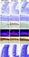
Neuronal apoptosis in Polβ-deficient mice is mediated by the p53-dependent pathway. Coronal sections of E13.5 telencephalons (A to D) and E11.5 spinal cords and dorsal root ganglions (E to H) in Polβ+/+ p53+/+ (wild-type) (A and E), Polβ−/− p53+/+ (B and F), Polβ−/− p53+/− (C and G), and Polβ−/− p53−/− (D and H) embryos were stained with anti-cleaved caspase-3 antibody. DRG, dorsal root ganglion; LGE, lateral ganglionic eminence; MGE, medial ganglionic eminence; NCX, neocortex; SC, spinal cord.
FIG. 4.
FIG. 4.
Neuronal progenitors in E13.5 Polβ−/− p53−/− embryos appear to normally proliferate and differentiate. Coronal sections of telencephalons in Polβ+/+ p53+/+ (wild-type) (A, D, G, J, M, and P), Polβ−/−p53+/+ (B, E, H, K, N, and Q), and Polβ−/− p53−/− (C, F, I, L, O, and R) embryos at E13.5 (A to O) and E14.5 (P to R) were stained with cresyl violet (A to F and P to R). The sections were also stained with anti-cleaved caspase-3 antibody (G to I), anti-PCNA antibody (red) and DAPI (blue) (J to L), or anti-neuron specific type III β-tubulin antibody TuJ1 (M to O). Arrows in panels A, C, P, Q, and R indicate the CP. Arrowheads in panel K indicate PCNA-activated cells. LV, lateral ventricle.
FIG. 5.
FIG. 5.
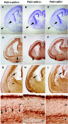
Development of the brain is incomplete in E18.5 Polβ−/− p53−/− embryos. Coronal sections of telencephalons in Polβ+/+ p53+/+ (wild-type) (A, D, G, and J), Polβ−/− p53+/+ (B, E, H, and K), and Polβ−/−p53−/− (C, F, I, and L) embryos at E18.5 were stained with cresyl violet (A to C), anti-phosphorylated neurofilament antibody SMI31 (D to I), and anticalbindin antibody (J to L). Photographs of the sections in each genotype were taken with the same magnification. The arrowheads in panels A, B, and C indicate part of the cytoarchitecture recovered moderately in Polβ−/− p53−/− mice compared to Polβ−/− p53+/+. The arrows in panels D, E, and F indicate the anterior commissure (AC). The arrows in panels G, H, and I indicate aberrant axonal tracts in the striatum (St). The arrows in panel J indicate calbindin-positive cells. MZ, marginal zone.

References

    1. Abe, H., O. Amano, T. Yamakuni, Y. Takahashi, and H. Kondo. 1990. Localization of spot 35-calbindin (rat cerebellar calbindin) in the anterior pituitary of the rat: developmental and sexual differences. Arch. Histol. Cytol. 53:585-591. - PubMed
    1. Anderson, S. A., D. D. Eisenstat, L. Shi, and J. L. Rubenstein. 1997. Interneuron migration from basal forebrain to neocortex: dependence on Dlx genes. Science 278:474-476. - PubMed
    1. Banasiak, K. J., and G. G. Haddad. 1998. Hypoxia-induced apoptosis: effect of hypoxic severity and role of p53 in neuronal cell death. Brain Res. 797:295-304. - PubMed
    1. Barnes, D. E., G. Stamp, I. Rosewell, A. Denzel, and T. Lindahl. 1998. Targeted disruption of the gene encoding DNA ligase IV leads to lethality in embryonic mice. Curr. Biol. 8:1395-1398. - PubMed
    1. Bayer, S. A., and J. Altman. 1991. Neocortical development. Raven Press, New York, N.Y.

VSports在线直播 - Publication types

VSports注册入口 - MeSH terms