Skip to main page content
U.S. flag

An official website of the United States government

Dot gov

The . gov means it’s official. Federal government websites often end in . gov or . mil. Before sharing sensitive information, make sure you’re on a federal government site VSports app下载. .

Https

The site is secure. The https:// ensures that you are connecting to the official website and that any information you provide is encrypted and transmitted securely V体育官网. .

. 2004 Sep 21;101(38):13850-5.
doi: 10.1073/pnas.0405146101. Epub 2004 Sep 13.

Disruption of ceruloplasmin and hephaestin in mice causes retinal iron overload and retinal degeneration with features of age-related macular degeneration (V体育官网)

Affiliations

VSports最新版本 - Disruption of ceruloplasmin and hephaestin in mice causes retinal iron overload and retinal degeneration with features of age-related macular degeneration

Paul Hahn et al. Proc Natl Acad Sci U S A. .

Abstract

Mechanisms of brain and retinal iron homeostasis have become subjects of increased interest after the discovery of elevated iron levels in brains of patients with Alzheimer's disease and retinas of patients with age-related macular degeneration. To determine whether the ferroxidase ceruloplasmin (Cp) and its homolog hephaestin (Heph) are important for retinal iron homeostasis, we studied retinas from mice deficient in Cp and/or Heph. In normal mice, Cp and Heph localize to Müller glia and retinal pigment epithelium, a blood-brain barrier. Mice deficient in both Cp and Heph, but not each individually, had a striking, age-dependent increase in retinal pigment epithelium and retinal iron. The iron storage protein ferritin was also increased in Cp-/-Heph-/Y retinas. After retinal iron levels had increased, Cp-/-Heph-/Y mice had age-dependent retinal pigment epithelium hypertrophy, hyperplasia and death, photoreceptor degeneration, and subretinal neovascularization, providing a model of some features of the human retinal diseases aceruloplasminemia and age-related macular degeneration. This pathology indicates that Cp and Heph are critical for CNS iron homeostasis and that loss of Cp and Heph in the mouse leads to age-dependent retinal neurodegeneration, providing a model that can be used to test the therapeutic efficacy of iron chelators and antiangiogenic agents VSports手机版. .


Figures (V体育平台登录)
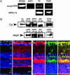
Fig. 1.
Fig. 1.
Cp and Heph mRNA and proteins are present in normal RPE and retina within Müller cells. (A) Bands from ethidium bromide-stained agarose gels corresponding to RT-PCR amplification products of the indicated mRNAs from dissected C57BL/6 murine RPE and cultured ARPE-19 cells. In all cases, the amplification product was of the expected size. (B) Western analysis of Cp and Heph in dissected C57BL/6 murine retinas (mRet) human retinas (huRet), cultured ARPE-19 cells, and cultured rMC-1 cells. Purified human Cp (labeled Cp) was used as control. (C) Control BALB/c retina labeled with a Cy3-conjugated donkey anti-rabbit secondary antibody. (D) Normal BALB/c retina immunolabeled for Heph. (E–G) Normal BALB/c retina double-labeled with anti-Heph and anti-CRALBP, a marker for Muller glia. A single red exposure reveals anti-Heph immunoreactivity (E). A single green exposure shows anti-CRALBP immunore-activity (F). Double exposure shows yellow colocalization of Heph and CRALBP (G). (Scale bars: 50 μm.)
Fig. 2.
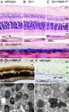
Fig. 2.
Adult (6-month-old) Cp–/–Heph–/Y RPE and photoreceptors accumulate iron. (A–C) 6-month-old WT (A), Cp–/– (B), and Cp–/–Heph–/Y (C) retinas Perls' stained for iron (blue) and counterstained with hematoxylin/eosin. (D) High magnification of Prussian blue Perls' label in 6-month-old Cp–/–Heph–/Y RPE. (E and F) Light photomicrographs of 6-month-old Cp–/–Heph–/Y (E) and WT (F) retinas after DAB enhancement (brown) of Perls' stain. (G–H) Electron micrographs of RPE from 6-month-old Cp–/–Heph–/Y (G) and WT (H) eyes. Only the Cp–/–Heph–/Y RPE (G) contains electrondense vesicles (*) sometimes fused with melanosomes.
Fig. 3.
Fig. 3.
Cp–/–Heph–/Y retinas have increased ferritin. Fluorescence photomicrographs of 6-month-old WT, Cp–/–, and Cp–/–Heph–/Y retinas immunolabeled for H-ferritin (A–C) and L-ferritin (D–F) and imaged by using identical exposure parameters. (Scale bars: 50 μm.)
Fig. 4.
Fig. 4.
Gross photomicrograph of atrophy in 9-month-old Cp–/–Heph–/Y retina. After removal of the cornea and lens, WT eye cup (A) has transillumination only at the optic nerve. In contrast, Cp–/–Heph–/Y eye cup (B) has areas of depigmentation (arrows) consistent with RPE pigment loss and atrophy in the peripheral retina.
Fig. 5.
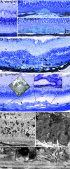
Fig. 5.
Nine-month-old Cp–/–Heph–/Y mice have retinal degeneration. (A) Light photomicrograph of WT retina. (B and C) Cp–/–Heph–/Y retina has focal patches of hypertrophic RPE cells in some areas (B) and confluent hypertrophic RPE cells in other areas (C). (D) In an area of RPE hyperplasia (demarcated by arrowheads), Cp–/–Heph–/Y retinas have local photoreceptor degeneration [demarcated by arrows in the outer nuclear layer (ONL)] and subretinal neovascularization (red *). (E) In an area of hypertrophic, hyperplastic (area demarcated by arrowheads) RPE cells, a necrotic RPE cell also observed by electron microscopy (Left Inset) is present. Within the area of RPE hyperplasia, there is local photoreceptor thinning and subretinal neovascularization (red *) visible as small vessels containing erythrocytes (Right Inset). The hyperplastic RPE have formed a localized cyst (Cy). (F) Electron micrograph of WT RPE. Br, Bruch's membrane; AM, apical microvilli; OS, photoreceptor outer segments. (G) Electron micrograph of Cp–/–Heph–/Y RPE overloaded with phagosomes and lysosomes containing photoreceptor outer segments at various stages of digestion. Some of these lysosomes (*) contained multilamellar structures characteristic of outer segment membranes (Inset). (H) Electron micrograph of Cp–/–Heph–/Y deposits between RPE and Bruch's membrane containing wide-spaced collagen (*). (Scale bars: AE, 50 μm; F and G, 2 μm; H, 500 nm.)

VSports在线直播 - References

    1. Doly, M., Bonhomme, B. & Vennat, J. C. (1986) Ophthalmic Res. 18, 21–27. - PubMed
    1. Vergara, O., Ogden, T. & Ryan, S. (1989) Exp. Eye Res. 49, 1115–1126. - PubMed
    1. Tawara, A. (1986) Invest. Ophthalmol. Visual Sci. 27, 226–236. - PubMed
    1. Hahn, P., Milam, A. H. & Dunaief, J. L. (2003) Arch. Ophthalmol. 121, 1099–1105. - V体育平台登录 - PubMed
    1. Miyajima, H., Nishimura, Y., Mizoguchi, K., Sakamoto, M., Shimizu, T. & Honda, N. (1987) Neurology 37, 761–767. - "V体育安卓版" PubMed

Publication types

"V体育官网入口" LinkOut - more resources