Skip to main page content
U.S. flag

An official website of the United States government

Dot gov

The . gov means it’s official. Federal government websites often end in VSports app下载. gov or . mil. Before sharing sensitive information, make sure you’re on a federal government site. .

Https

The site is secure. The https:// ensures that you are connecting to the official website and that any information you provide is encrypted and transmitted securely V体育官网. .

. 2004 Sep;24(17):7654-68.
doi: 10.1128/MCB.24.17.7654-7668.2004.

Ribosomal protein L23 activates p53 by inhibiting MDM2 function in response to ribosomal perturbation but not to translation inhibition

Affiliations

Ribosomal protein L23 activates p53 by inhibiting MDM2 function in response to ribosomal perturbation but not to translation inhibition

VSports - Mu-Shui Dai et al. Mol Cell Biol. 2004 Sep.

Abstract

The p53-MDM2 feedback loop is vital for cell growth control and is subjected to multiple regulations in response to various stress signals VSports手机版. Here we report another regulator of this loop. Using an immunoaffinity method, we purified an MDM2-associated protein complex that contains the ribosomal protein L23. L23 interacted with MDM2, forming a complex independent of the 80S ribosome and polysome. The interaction of L23 with MDM2 was enhanced by treatment with actinomycin D but not by gamma-irradiation, leading to p53 activation. This activation was inhibited by small interfering RNA against L23. Ectopic expression of L23 reduced MDM2-mediated p53 ubiquitination and also induced p53 activity and G(1) arrest in p53-proficient U2OS cells but not in p53-deficient Saos-2 cells. These results reveal that L23 is another regulator of the p53-MDM2 feedback regulation. .


Figures

FIG. 1.
FIG. 1.
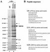
Isolation of a human MDM2-associated protein complex by immunoaffinity purification. (A) Colloidal blue staining analysis of proteins eluted from 12CA5 beads loaded with either empty vector expressing 293 cytoplasmic extracts (293) or the cytoplasmic extracts from the 293-HA-MDM2 cell line (lane 2). MDM2-associated polypeptides were digested and subjected to sequence analysis by mass spectrometry. The MDM2, L5, L11, and L23 bands are indicated. The question marks denote unidentified polypeptides. α-HA, anti-HA. (B) Peptide sequences for L5, L11, and L23 bands obtained from mass spectrometry analysis.
FIG. 2.
FIG. 2.
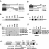
L23 interacts with MDM2 in cells. (A) Exogenous MDM2 and L23 interact with each other in 293 cells. HA-MDM2 (1.5 μg), Flag-L23 (1.5 μg), or both vectors (1.5 μg each) were used for transfection, as indicated at the top. Whole-cell lysates (500 μg) were subjected to immunoprecipitation (IP) with anti-HA (α-HA) or a control antibody followed by immunoblotting (IB) with anti-Flag (α-Flag) (upper) or anti-HA (lower) antibodies. IgG, immunoglobulin G. (B) The same transfections as shown in panel A were conducted except that anti-Flag antibodies were used for immunoprecipitation. (C) MDM2 colocalized with L23 in both the nucleus and the cytoplasm but not in the nucleolus. 293-HA-MDM2 cells were transfected with Flag-L23 and immunostained with both polyclonal anti-MDM2 (red) and monoclonal anti-Flag (green) antibodies. (D) L23 binds to MDM2 deletion mutants in cells. 293 cells were transfected with 6 μg of wild-type MDM2, MDM2Δ150-230, or empty (−) plasmids, as indicated at the top. Whole-cell lysates (500 μg) were immunoprecipitated with anti-L23 (α-L23) antibodies followed by immunoblotting with anti-MDM2 (2A10) and anti-L23 antibodies. Ten percent of the lysates loaded as input are shown in the panels on the right side. (E) Endogenous L23 interacts with endogenous MDM2 in U2OS cells. Whole-cell lysate (500 μg) was used for immunoprecipitation with either rabbit polyclonal anti-L23 antibody or preimmune serum (control), followed by immunoblotting with anti-MDM2 (2A10) (top panel) or anti-L23 (bottom panel) antibody.
FIG. 3.
FIG. 3.
Ribosomal protein L23 interacts with MDM2 in vitro. (A) Schematic presentation of recombinant full-length MDM2 and its fragments fused to GST. Black rectangles indicate GST. Gray rectangles indicate MDM2 or its fragments. (B) L23 preferentially binds to the acidic domains of MDM2. About 200 ng of purified GST alone, full-length GST-MDM2, or GST-MDM2 deletion mutants including MDM2/1-150, MDM2/1-301, MDM2/294-491, MDM2/384-491, and MDM2/425-491 immobilized on glutathione beads were used in GST pull-down assays with 200 ng of His-L23 purified from bacteria. Bound L23 was detected by immunoblotting (IB) with anti-L23 (α-L23) antibodies. (C) Coomassie blue staining of GST-MDM2 fusion proteins used in panel B. (D) Schematic presentation of recombinant full-length L23 and its fragments fused to GST. (E) MDM2 binds to the middle domain of L23 in vitro. The same GST fusion protein pull-down assay as that described for panel B was conducted except that purified 200 ng of GST-L23 and 200 ng of GST-L23 deletion mutants were incubated with 200 ng of His-MDM2 purified from bacteria, as indicated. Bound MDM2 was detected by immunoblotting with anti-MDM2 antibodies (2A10). (F) Immunoblot of GST-L23 fusion proteins used in panel E with anti-L23 antibodies. (G) Schematic presentation of MDM2 domains that bind to L23, L5, and L11.
FIG. 4.
FIG. 4.
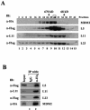
MDM2 forms a complex with L23, L5, and L11 in cells. (A) MDM2 cosedimented with L23, L5, and L11. Whole-cell lysates (2 mg) prepared from HA-MDM2-expressing 293 cells were subjected to a 12.5 to 25% glycerol gradient sedimentation centrifugation. Fractions were collected for immunoblot (IB) analysis with antibodies indicated to the left of each panel. Molecular markers coeluted with fractions are indicated at the top. Fraction 30, boxed, was used for immunoprecipitation and immunoblot analysis shown in panel B. (B) MDM2 is coimmunoprecipitated with L23, L5, and L11 in fraction 30. Fraction 30 (200 μl) was used for immunoprecipitation (IP) with the anti-HA (α-HA) or control antibody as indicated at the top, followed by immunoblotting with the antibodies indicated to the left. IgG, immunoglobulin G; α-Flag, anti-Flag; α-L11, anti-L11.
FIG. 5.
FIG. 5.
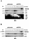
MDM2 does not associate with the 80S ribosome and polysomes. (A) Ectopically expressed MDM2 does not associate with polysomes. Cytoplasmic extracts (5 mg) containing polysomes from HA-MDM2-expressing 293 cells were subjected to a 15 to 47% linear sucrose gradient sedimentation centrifugation. Fourteen fractions were collected, and 30 μl of each fraction was used for immunoblotting (IB) with anti-HA (α-HA), anti-L11 (α-L11), or anti-L23 (α-L23) antibodies as indicated to the left. Total RNAs were isolated from each fraction and subjected to electrophoresis on a 1% agarose gel and stained with ethidium bromide as shown in the bottom panel. 28S, 18S, 5.8S, and 5S rRNAs are indicated to the right. The fractions containing polysomes and mRNPs are indicated on the top. The 80S ribosome is indicated at the bottom. (B) Endogenous MDM2 does not associate with polysomes. The same fractionation as that shown in panel A was performed with U2OS cell extracts. The distributions of polysomes and mRNPs are indicated. Thirty microliters of each fraction was subjected to immunoblot analysis with anti-MDM2 (2A10, α-MDM2), anti-L11, or anti-L23 antibodies as indicated to the left.
FIG. 6.
FIG. 6.
Ectopic expression of L23 stabilizes p53 and inhibits MDM2-mediated p53 ubiquitination. (A) Ectopic expression of L23 reverses MDM2-mediated p53 degradation. H1299 cells were transfected with 1.0 μg of Flag-L23 in the presence of p53 (0.5 μg) with (+) or without (−) MDM2 (1.0 μg) as indicated. Cell lysates (50 μg) were immunoblotted (IB) with anti-MDM2 (α-MDM2), anti-p53 (α-p53), anti-Flag (α-Flag), or antitubulin (α-tubulin) antibodies as indicated to the left. (B) L23 inhibits MDM2-mediated p53 ubiquitination in cells. H1299 cells were transfected with combinations of L23 (2 μg)-, p53 (1 μg)-, or MDM2 (1 μg)-encoding plasmids in the presence of the His-ubiquitin (His-Ub) (2 μg) plasmid as indicated at the top. The cells were treated with MG132 (20 μM) for 8 h before harvesting. The in vivo ubiquitination assay was performed as described in Materials and Methods. Ubiquitinated proteins were detected by immunoblotting with the anti-p53 (DO-1) and anti-HA (α-HA) antibodies. Ubiquitinated p53s [p53-(His-Ub)n] and ubiquitinated MDM2s [MDM2-(His-Ub)n] are indicated to the right of the upper and middle panels. The expression levels of MDM2, p53, and L23 are shown in the lower panels. (C) Ectopic expression of L23 induces endogenous p53. U2OS cells were transfected with 1.0 μg (lane 2) or 2.0 μg (lane 3) of Flag-L23 or 2.0 μg of Flag-L23ΔN (lane 4) plasmids. Cell lysates (50 μg) were used for immunoblot analysis with antibodies as indicated to the right. α-p21, anti-p21.
FIG. 7.
FIG. 7.
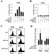
Ectopic expression of L23 stimulates p53-dependent transcription and G1 arrest. (A) Ectopic expression of L23 increases p53RE-dependent luciferase activity in p53-proficient U2OS cells. U2OS cells were transfected with increasing amounts of Flag-L23 (0.4 μg [1×] and 0.8 μg [2×]) or Flag-L23ΔN (0.4 μg [1×] and 0.8 μg [2×]) in the presence of a luciferase reporter plasmid driven by p53RE (BP100 luc, 0.1 μg) or a control luciferase reporter plasmid (TB luc, 0.1 μg). Luciferase activity is presented in arbitrary units. (B) L23 does not affect p53RE-dependent luciferase activity in p53-deficient Saos-2 cells. The same transfection followed by a luciferase assay as that described for panel A was conducted except Saos-2 cells were used here. (C) Ectopic expression of L23 leads to p53-dependent G1 cell cycle arrest. U2OS or Saos-2 cells were transfected with GFP (2 μg), GFP-L23 (2 μg), or GFP-L23ΔC (2 μg) plasmids and treated with nocodazole as described in Materials and Methods. GFP-expressing cells were then gated for cell cycle analysis. The histograms of PI staining from one representative experiment are shown. Percentages indicate the cells that were arrested in G1 phase. (D) The mean percentage of cells arrested in the G1 phase obtained from four separate experiments is presented. Bars indicate standard deviations. +, present; −, absent.
FIG. 8.
FIG. 8.
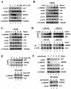
A low dose of actinomycin D enhances MDM2-L23 interaction and p53 activation. (A) Low doses of actinomycin D induce p53 and its function, whereas high doses of actinomycin D induce p53 but not its function. U2OS cells were treated with increasing amounts of actinomycin D (Act D) as indicated at the top. Cell lysates (50 μg) were used for an immunoblot analysis with antibodies indicated to the right. (B) Time-dependent effect of actinomycin D on p53 and L23 levels in U2OS cells. U2OS cells were treated with 5 nM actinomycin D and harvested at different time points as indicated at the top. Cell lysates (50 μg) were used for an immunoblot analysis with antibodies indicated to the left of each panel. (C) Time-dependent effect of actinomycin D on p53 and L23 levels in WI38 cells. WI38 cells were treated with 5 nM actinomycin D, harvested, and blotted with antibodies as described for panel B. (D) Five nanomolar actinomycin D enhances MDM2-L23 interaction. U2OS cells were treated with 5 nM actinomycin D and harvested at different time points as indicated at the top. Cells were incubated with (+) or without (−) MG132 (20 μM) for 6 h before harvesting. Cell lysates (500 μg) were subjected to an immunoprecipitation (IP) with anti-L23 antibodies and immunoblotting with anti-MDM2 or anti-L23 antibodies (lower panels). The lysates were also directly loaded onto an SDS gel for an immunoblot analysis with anti-MDM2, anti-p53, or anti-L23 antibodies (upper panels). (E) Ionizing irradiation does not affect L23-MDM2 interaction. U2OS cells were treated with gamma irradiation (10 Gy) and harvested at different time points as indicated. Cell lysates (50 μg) were subjected to an immunoblot analysis with anti-p53, anti-MDM2, or anti-L23 antibodies (upper panels). The cell lysates (500 μg) were subjected to an immunoprecipitation analysis with anti-L23 antibodies, followed by an immunoblot analysis with anti-MDM2 or anti-L23 antibodies (bottom panels). (F) Pactamycin treatment does not induce p53 and the MDM2-L23 interaction. U2OS cells were treated with 0.2 μg of pactamycin/ml for different numbers of hours as indicated at the top. The cells were harvested for immunoblotting with the indicated antibodies (top panels). The cell lysates were also subjected to immunoprecipitation with anti-L23 followed by immunoblotting with anti-MDM2 and anti-L23 antibodies (bottom panels). α-L23, anti-L23; α-p53, anti-p53; α-MDM2, anti-MDM2; α-p21, anti-p21; α-tubulin, antitubulin; α-L11, anti-L11.
FIG. 9.
FIG. 9.
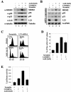
Inhibition of endogenous L23 by siRNA induces p53 but inhibits actinomycin D (Act D)-induced p53 activation and G1 arrest. (A) Inhibition of endogenous L23 by siRNA induces the p53 level but inhibits actinomycin D-induced p53 induction. U2OS cells were transfected with L23 siRNA oligonucleotides (0.2 μM, lanes 2 and 4) or a scrambled RNA duplex (0.2 μM, lanes 1 and 3). Cells were then incubated with (+) (lanes 3 and 4) or without (−) (lanes 1 and 2) 5 nM actinomycin D for 8 h before harvesting. Cell lysates (50 μg) were then immunoblotted with anti-MDM2, anti-p53, anti-p21, anti-L23, or antitubulin antibodies. (B) Inhibition of endogenous L23 by siRNA inhibits actinomycin D-induced p53 transcriptional activity. Cells were prepared as described for panel A, and total RNA was extracted. RT-PCR analysis was performed to detect MDM2, p53, p21, L23, and GAPDH mRNA levels as indicated. (C) Actinomycin-induced G1 arrest is inhibited by elimination of endogenous L23 by siRNA. U2OS cells were treated as described for panel A except that cells were treated with nocodazole for 16 h before harvesting. Cells were then stained with PI for fluorescence-activated cell sorter analysis. The histograms of PI staining from the results of one representative experiment are shown. (D) Summary of the results from three independent experiments as described for panel C. The mean percentages of cells arrested in G1 phase are presented. Bars indicate standard deviations. (E) Elimination of endogenous L23 by siRNA inhibits actinomycin D-induced p53RE-dependent luciferase activity. U2OS cells were transfected with a luciferase reporter plasmid driven by the p53RE (BP100 luc, 0.2 μg) and β-galactosidase (0.2 μg) plasmids followed by treatment with L23 siRNA and actinomycin D as described for panel A. Luciferase activity is presented in arbitrary units. α-L23, anti-L23; α-p53, anti-p53; α-MDM2, anti-MDM2; α-p21, anti-p21; α-tubulin, antitubulin; DMSO, dimethyl sulfoxide.
FIG. 10.
FIG. 10.
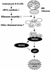
A model for p53 activation by interfering with the MDM2-p53 feedback loop in response to perturbation of ribosomal biogenesis. Bars indicate inhibition, and arrows indicate activation.

References

    1. Ashcroft, M., M. H. Kubbutat, and K. H. Vousden. 1999. Regulation of p53 function and stability by phosphorylation. Mol. Cell. Biol. 19:1751-1758. - PMC - PubMed
    1. Ashcroft, M., Y. Taya, and K. H. Vousden. 2000. Stress signals utilize multiple pathways to stabilize p53. Mol. Cell. Biol. 20:3224-3233. - PMC - PubMed
    1. Banin, S., L. Moyal, S. Shieh, Y. Taya, C. W. Anderson, L. Chessa, N. I. Smorodinsky, C. Prives, Y. Reiss, Y. Shiloh, and Y. Ziv. 1998. Enhanced phosphorylation of p53 by ATM in response to DNA damage. Science 281:1674-1677. - PubMed
    1. Barak, Y., T. Juven, R. Haffner, and M. Oren. 1993. mdm2 expression is induced by wild type p53 activity. EMBO J. 12:461-468. - PMC - PubMed
    1. Boyd, M. T., N. Vlatkovic, and D. S. Haines. 2000. A novel cellular protein (MTBP) binds to MDM2 and induces a G1 arrest that is suppressed by MDM2. J. Biol. Chem. 275:31883-31890. - PubMed

Publication types (VSports手机版)

MeSH terms