Skip to main page content
U.S. flag

An official website of the United States government

Dot gov

The VSports app下载. gov means it’s official. Federal government websites often end in . gov or . mil. Before sharing sensitive information, make sure you’re on a federal government site. .

Https

The site is secure. The https:// ensures that you are connecting to the official website and that any information you provide is encrypted and transmitted securely V体育官网. .

. 2004 Aug;24(15):6763-72.
doi: 10.1128/MCB.24.15.6763-6772.2004.

Reactive nitrogen species-induced cell death requires Fas-dependent activation of c-Jun N-terminal kinase

Affiliations

Reactive nitrogen species-induced cell death requires Fas-dependent activation of c-Jun N-terminal kinase

Punya Shrivastava et al. Mol Cell Biol. 2004 Aug.

Abstract

Nitrogen dioxide is a highly toxic reactive nitrogen species (RNS) recently discovered as an inflammatory oxidant with great potential to damage tissues. We demonstrate here that cell death by RNS was caused by c-Jun N-terminal kinase (JNK). Activation of JNK by RNS was density dependent and caused mitochondrial depolarization and nuclear condensation. JNK activation by RNS was abolished in cells lacking functional Fas or following expression of a truncated version of Fas lacking the intracellular death domain VSports手机版. In contrast, RNS induced JNK potently in cells expressing a truncated version of tumor necrosis factor receptor 1 or cells lacking tumor necrosis factor receptor 1 (TNF-R1), illustrating a dependence of Fas but not TNF-R1 in RNS-induced signaling to JNK. Furthermore, Fas was oxidized, redistributed, and colocalized with Fas-associated death domain (FADD) in RNS-exposed cells, illustrating that RNS directly targeted Fas. JNK activation and cell death by RNS occurred in a Fas ligand- and caspase-independent manner. While the activation of JNK by RNS or FasL required FADD, the cysteine-rich domain 1 containing preligand assembly domain required for FasL signaling was not involved in JNK activation by RNS. These findings illustrate that RNS cause cell death in a Fas- and JNK-dependent manner and that this occurs through a pathway distinct from FasL. Thus, avenues aimed at preventing the interaction of RNS with Fas may attenuate tissue damage characteristic of chronic inflammatory diseases that are accompanied by high levels of RNS. .


Figures
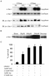
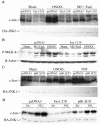
FIG. 1.
FIG. 1.
RNS-induced JNK activation in lung epithelial cells. (A) Log-phase or confluent cultures of RLE cells were exposed to 10 ppm of NO2 for 4 h. Extracts prepared at different times were examined either for JNK activation via an in vitro kinase assay (upper panel) or phosphorylation of c-Jun (lower panel) by Western blotting (immunoblotting [IB]). (B) Log-phase cultures of C10 cells were exposed to different concentrations of ONOO for 2 h, and JNK activity was evaluated. Decomposed ONOO (Decomp) was used as a reagent control. (C) C10 cells were preincubated with N-acetyl-l-cysteine (NAC; pH 7.4) or ascorbate (Asc) at the indicated concentrations for 15 min prior to exposure to 200 μM ONOO for 2 h. Cells were harvested for assessment of viability with the MTT assay. Results are expressed as percent viability of sham controls. *, P < 0.05 (analysis of variance) compared to ONOO-treated cells; #, P < 0.05 (analysis of variance) compared to ONOO-treated cells, but not different from sham controls.
FIG. 2.
FIG. 2.
Expression of JNK1-APF inhibits RNS-induced cell death. (A) Assessment of JNK activation and c-Jun phosphorylation in pcDNA3 or JNK1-APF transfected cell pools. Cells were treated with 200 μM ONOO for 2 h for assessment of JNK activation in an in vitro kinase assay (top) or c-Jun phosphorylation by Western blotting (bottom). (Middle panel) Western blotting for JNK1. The expression of JNK1-APF is indicated. pcDNA3 or JNK1-APF cells grown on glass coverslips were treated with 200 μM ONOO for the indicated times for the assessment of mitochondrial membrane potential (B) or nuclear condensation (C). Mitochondrial membrane potential was determined by incubation with 5-μg/ml JC-1. Polarized mitochondria are visualized by punctate red staining, whereas depolarized mitochondria are visualized by their diffuse green staining. Cells were counted in five random fields, and data are expressed as a percentage of polarized cells. Nuclear condensation was determined after staining of cells with 5-μg/ml Hoechst. Cells were counted in five random fields, and data are expressed as a percentage of cells containing intact nuclei. Results are expressed as means ± standard error. *, P < 0.05 compared to sham controls; #, P < 0.05 compared to pcDNA3 vector controls (analysis of variance). (D) Assessment of AIF nuclear localization in cells exposed to 50 nM EPO in the presence of 15-mU/ml glucose oxidase (GO) and 100 μM sodium nitrite (NO2) for 4 h. Nuclear presence of AIF (green) was confirmed via confocal microscopy and colocalization with propidium iodide (blue). (E) Assessment of viability of C57BL/6 or JNK1−/− lung fibroblasts exposed to 200 μM ONOO, using the MTT assay. Data are expressed as percent survival compared to sham controls. *, P < 0.05 (analysis of variance) compared to C57BL/6-derived fibroblasts exposed to ONOO for 1 h; #, P < 0.05 (analysis of variance) compared to C57BL/6-derived fibroblasts exposed to ONOO for 2 h.
FIG. 2.
FIG. 2.
Expression of JNK1-APF inhibits RNS-induced cell death. (A) Assessment of JNK activation and c-Jun phosphorylation in pcDNA3 or JNK1-APF transfected cell pools. Cells were treated with 200 μM ONOO for 2 h for assessment of JNK activation in an in vitro kinase assay (top) or c-Jun phosphorylation by Western blotting (bottom). (Middle panel) Western blotting for JNK1. The expression of JNK1-APF is indicated. pcDNA3 or JNK1-APF cells grown on glass coverslips were treated with 200 μM ONOO for the indicated times for the assessment of mitochondrial membrane potential (B) or nuclear condensation (C). Mitochondrial membrane potential was determined by incubation with 5-μg/ml JC-1. Polarized mitochondria are visualized by punctate red staining, whereas depolarized mitochondria are visualized by their diffuse green staining. Cells were counted in five random fields, and data are expressed as a percentage of polarized cells. Nuclear condensation was determined after staining of cells with 5-μg/ml Hoechst. Cells were counted in five random fields, and data are expressed as a percentage of cells containing intact nuclei. Results are expressed as means ± standard error. *, P < 0.05 compared to sham controls; #, P < 0.05 compared to pcDNA3 vector controls (analysis of variance). (D) Assessment of AIF nuclear localization in cells exposed to 50 nM EPO in the presence of 15-mU/ml glucose oxidase (GO) and 100 μM sodium nitrite (NO2) for 4 h. Nuclear presence of AIF (green) was confirmed via confocal microscopy and colocalization with propidium iodide (blue). (E) Assessment of viability of C57BL/6 or JNK1−/− lung fibroblasts exposed to 200 μM ONOO, using the MTT assay. Data are expressed as percent survival compared to sham controls. *, P < 0.05 (analysis of variance) compared to C57BL/6-derived fibroblasts exposed to ONOO for 1 h; #, P < 0.05 (analysis of variance) compared to C57BL/6-derived fibroblasts exposed to ONOO for 2 h.
FIG. 3.
FIG. 3.
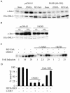
RNS-induced JNK activation occurs in Fas-dependent but TNF-R1-independent manner. (A) C10 cells were transfected with pcDNA3 or Fas 1-210 in the presence of HA-JNK1 (0.4 μg) and then exposed to 200 μM ONOO for 2 h or 1-μg/ml M2 plus 100-ng/ml FasL (30 min). HA-JNK was immunoprecipitated, and JNK activity was assayed by in vitro kinase assay. Levels of HA-JNK1 (bottom) are included as a loading control. (B) Cells were transfected with pcDNA3 or Fas 1-210, treated with agents as described in panel A, prior to assessment of phospho-MKK4 (top) by Western blotting. (Bottom) β-Actin expression. (C) C10 cells were transfected with pcDNA3 or p60ΔCD (1.6 μg) in the presence of HA-JNK1 (0.4 μg) and then exposed to ONOO as described above or to 10-ng/ml TNF-α (15 min) prior to assessment of JNK activity. Levels of HA-JNK1 are included as a loading control. (D) C10 cells were transfected with pcDNA3, Fas 1-210, or p60ΔCD (1.6 μg) in the presence of HA-JNK1 (0.4 μg) and then exposed to 40 ppm of NO2 for 2 h for assessment of JNK activity. Levels of HA-JNK1 are included as a loading control. (E) Lung fibroblasts derived from wild-type C57BL/6, LPR, or TNF-R1−/− mice were exposed to ONOO, FasL + M2, or TNF-α as described above, and JNK activity was assessed in an in vitro kinase assay (top). The bottom panel represents a portion of a JNK1 Western blot as a loading control. (F) Assessment of viability of C57BL/6 or LPR lung fibroblasts exposed to 200 μM ONOO for 2 h, using the MTT assay. Data are expressed as percent survival compared to sham controls. *, P < 0.05 (Student's t test) compared to C57BL/6-derived ONOO-exposed fibroblasts.
FIG. 3.
FIG. 3.
RNS-induced JNK activation occurs in Fas-dependent but TNF-R1-independent manner. (A) C10 cells were transfected with pcDNA3 or Fas 1-210 in the presence of HA-JNK1 (0.4 μg) and then exposed to 200 μM ONOO for 2 h or 1-μg/ml M2 plus 100-ng/ml FasL (30 min). HA-JNK was immunoprecipitated, and JNK activity was assayed by in vitro kinase assay. Levels of HA-JNK1 (bottom) are included as a loading control. (B) Cells were transfected with pcDNA3 or Fas 1-210, treated with agents as described in panel A, prior to assessment of phospho-MKK4 (top) by Western blotting. (Bottom) β-Actin expression. (C) C10 cells were transfected with pcDNA3 or p60ΔCD (1.6 μg) in the presence of HA-JNK1 (0.4 μg) and then exposed to ONOO as described above or to 10-ng/ml TNF-α (15 min) prior to assessment of JNK activity. Levels of HA-JNK1 are included as a loading control. (D) C10 cells were transfected with pcDNA3, Fas 1-210, or p60ΔCD (1.6 μg) in the presence of HA-JNK1 (0.4 μg) and then exposed to 40 ppm of NO2 for 2 h for assessment of JNK activity. Levels of HA-JNK1 are included as a loading control. (E) Lung fibroblasts derived from wild-type C57BL/6, LPR, or TNF-R1−/− mice were exposed to ONOO, FasL + M2, or TNF-α as described above, and JNK activity was assessed in an in vitro kinase assay (top). The bottom panel represents a portion of a JNK1 Western blot as a loading control. (F) Assessment of viability of C57BL/6 or LPR lung fibroblasts exposed to 200 μM ONOO for 2 h, using the MTT assay. Data are expressed as percent survival compared to sham controls. *, P < 0.05 (Student's t test) compared to C57BL/6-derived ONOO-exposed fibroblasts.
FIG. 4.
FIG. 4.
RNS-mediated activation of JNK does not require new protein synthesis and occurs in a FasL-independent manner. C10 cells were exposed to 200 μM ONOO for 15 min and then chased for 2 h in the presence or absence of 10 μM CHX (A) or with 10-μg/ml anti-FasL antibody (B) or 250-ng/ml Fas-Fc (C). JNK activity was assayed via an in vitro kinase assay. In control experiments, cells were treated with 1-μg/ml M2 plus 100-ng/ml FasL (30 min) in the presence of either anti-FasL antibody (B) or Fas-Fc (C).
FIG. 5.
FIG. 5.
RNS-dependent activation of JNK occurs in FADD-dependent but caspase-independent manner. C10 cells were transfected with either pcDNA3 or FADD (80-205) (1.6 μg) in the presence of HA-JNK1 (0.4 μg) and then exposed to 200 μM ONOO for 2 h (A) or 40 ppm of NO2 for 2 h (B). HA-JNK was immunoprecipitated, and JNK activity was assayed as described before. (C) C10 cells pretreated with 10 μM zVAD or IETD (30 min) were exposed to 200 μM ONOO (2 h) or 1-μg/ml M2 plus 100-ng/ml FasL (30 min) prior to assessment of JNK activity. (D) ONOO-induced cell death is caspase independent. C10 cells were pretreated with 10 μM zVAD or IETD (30 min) prior to exposure to 200 μM ONOO (2 h) or 1-μg/ml M2 plus 100-ng/ml FasL (1 h) prior to assessment of cell death by using the MTT assay.
FIG. 6.
FIG. 6.
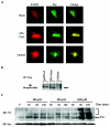
RNS cause aggregation and oxidative modification of Fas. (A) Cells grown on glass coverslips were treated with 200 μM ONOO or 1-μg/ml M2 plus 100-ng/ml FasL for 5 min, subsequently stained with anti-Fas or anti-FADD antibodies followed by fluorophore-conjugated secondary antibodies. Fas (green), FADD (red), and their colocalization (yellow) were examined by confocal microscopy. (B) C10 cells were treated with either 200 μM ONOO or M2 + FasL for 5 min, lysed in buffer containing 50 μM of MPB. Fas was immunoprecipitated (IP), and the extent of biotinylation was determined by using streptavidin peroxidase by Western blotting (immunoblotting [IB]). (C) C10 cells were treated with different concentrations of ONOO for indicated times. Fas was immunoprecipitated, and its nitrotyrosine content was evaluated by Western blotting. Fas monomer is indicated by the arrow, whereas higher-molecular-weight complexes are indicated by the bracket.
FIG. 7.
FIG. 7.
ONOO requires CRD2 and/or CRD3 but not CRD1 of Fas for signaling to JNK. (A) Schematic representation of Fas receptor. (B) C10 cells were transfected with 2 μg of the indicated constructs, and expression was confirmed by probing Western blots (immunoblots [IB]) with antibody directed against HA. Arrows represent the expected proteins. ns, nonspecific reactivity. (C) C10 cells were transfected with either pcDNA3, Fas 1-210, or Fas 67-210 in the presence of HA-JNK1 and then exposed to ONOO or FasL + M2 for assessment of JNK activity, as described before. Levels of HA-JNK1 are included as a loading control.

References

    1. Augusto, O., M. G. Bonini, A. M. Amanso, E. Linares, C. C. Santos, and S. L. De Menezes. 2002. Nitrogen dioxide and carbonate radical anion: two emerging radicals in biology. Free Radic. Biol. Med. 32:841-859. - PubMed
    1. Aulak, K. S., M. Miyagi, L. Yan, K. A. West, D. Massillon, J. W. Crabb, and D. J. Stuehr. 2001. Proteomic method identifies proteins nitrated in vivo during inflammatory challenge. Proc. Natl. Acad. Sci. USA 98:12056-12061. - PMC - PubMed
    1. Behrens, A., M. Sibilia, and E. F. Wagner. 1999. Amino-terminal phosphorylation of c-Jun regulates stress-induced apoptosis and cellular proliferation. Nat. Genet. 21:326-329. - PubMed
    1. Brennan, M. L., W. Wu, X. Fu, Z. Shen, W. Song, H. Frost, C. Vadseth, L. Narine, E. Lenkiewicz, M. T. Borchers, A. J. Lusis, J. J. Lee, N. A. Lee, H. M. Abu-Soud, H. Ischiropoulos, and S. L. Hazen. 2002. A Tale of Two Controversies. Defining both the role of peroxidases in nitrotyrosine formation in vivo using eosinophil peroxidase and myeloperoxidase-deficient mice, and the nature of peroxidase-generated reactive nitrogen species. J. Biol. Chem. 277:17415-17427. - PubMed
    1. Butterfield, L., B. Storey, L. Maas, and L. E. Heasley. 1997. c-Jun NH2-terminal kinase regulation of the apoptotic response of small cell lung cancer cells to ultraviolet radiation. J. Biol. Chem. 272:10110-10116. - PubMed (V体育2025版)

VSports最新版本 - Publication types

V体育平台登录 - MeSH terms

Substances